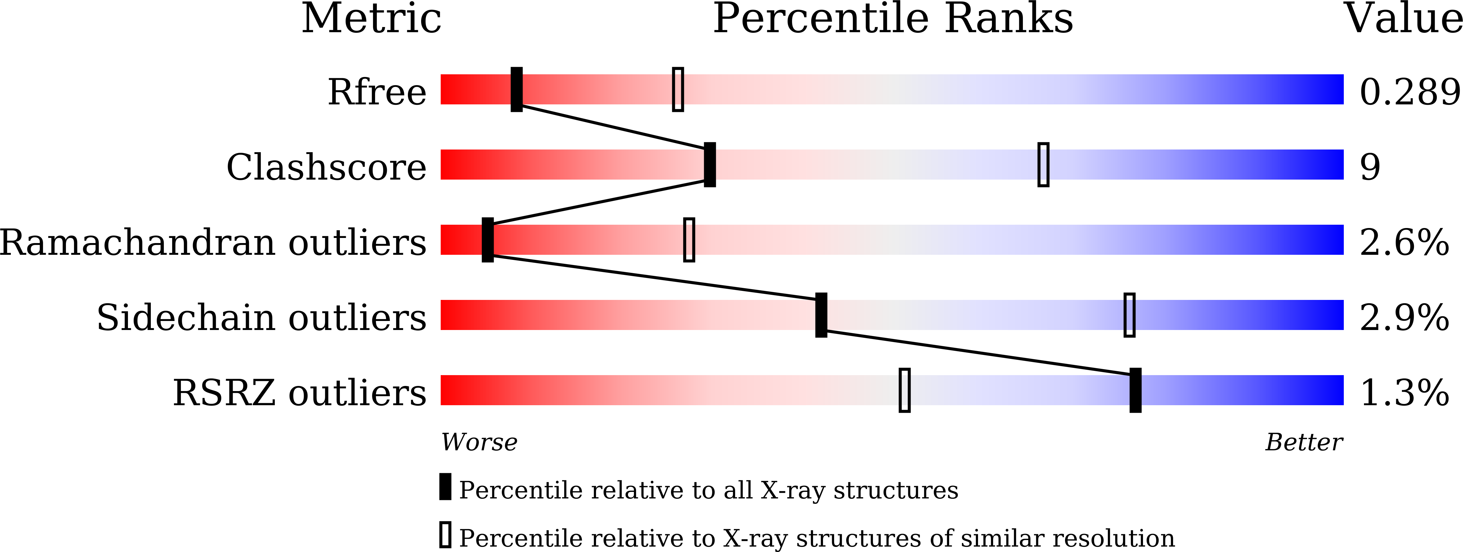
7F5Z image
Deposition Date 2021-06-23
Release Date 2022-05-11
Last Version Date 2023-11-29
Entry Detail
PDB ID:
7F5Z
Title:
Crystal structure of the single-stranded dna-binding protein from Mycobacterium tuberculosis- Form III
Biological Source:
Method Details:
Experimental Method:
Resolution:
3.00 Å
R-Value Free:
0.28
R-Value Work:
0.24
R-Value Observed:
0.25
Space Group:
P 61 2 2
Macromolecular Entities
Structures with similar UniProt ID
Protein Blast
Polymer Type:polypeptide(L)
Molecule:Single-stranded DNA-binding protein
Gene (Uniprot):ssb
Chain IDs:A, B
Chain Length:164
Number of Molecules:2
Biological Source:Mycobacterium tuberculosis (strain ATCC 25618 / H37Rv)
Primary Citation
Structural variability of Mycobacterium tuberculosis SSB and susceptibility to inhibition.
Curr.Sci. 122 281 289 (2022)
DOI: 10.18520/cs/v122/i3/281-289
Primary Citation of related structures