Deposition Date
2021-06-23
Release Date
2022-05-11
Last Version Date
2023-11-29
Entry Detail
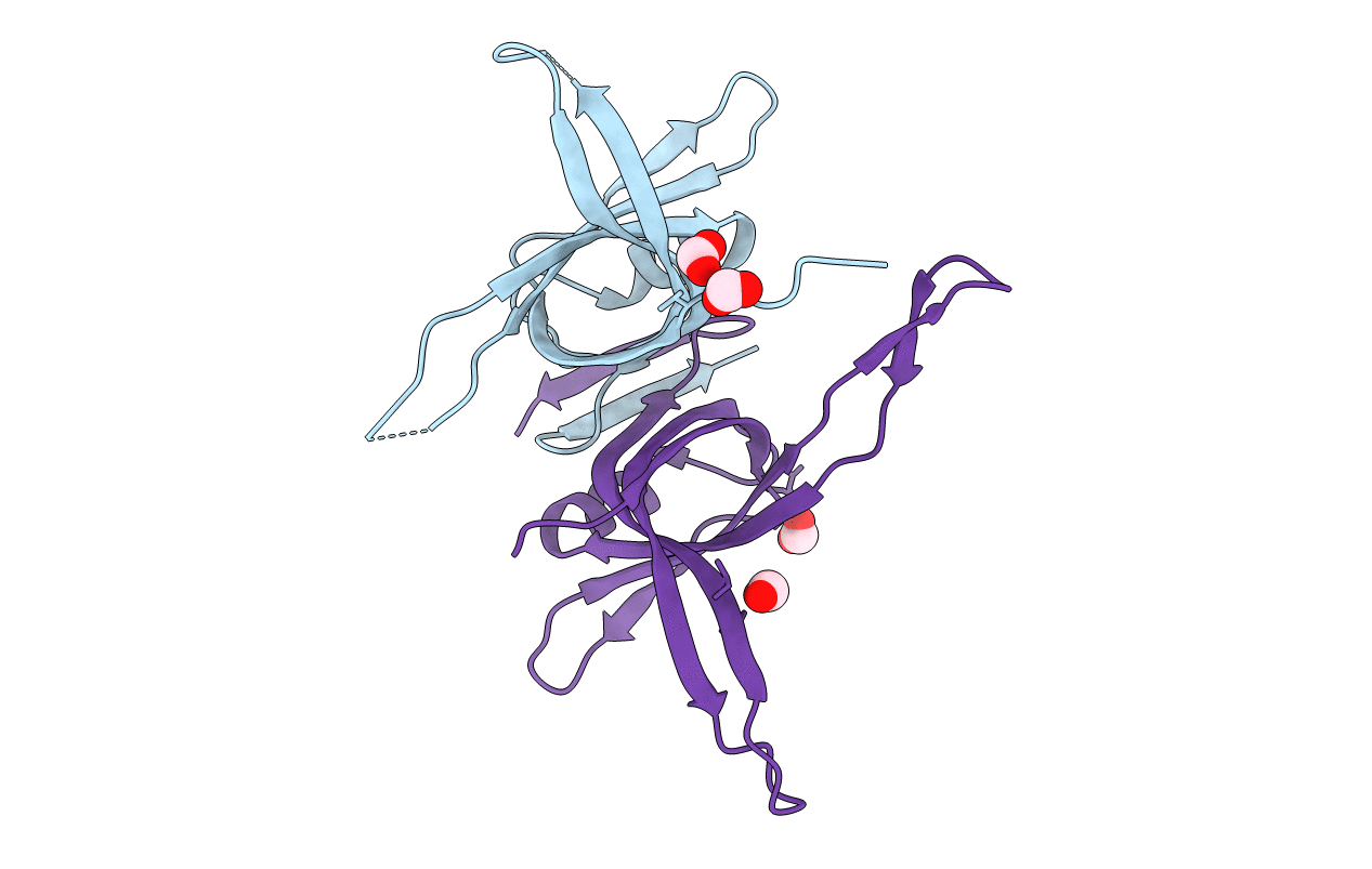
PDB ID:
7F5Y
Keywords:
Title:
Crystal structure of the single-stranded dna-binding protein from Mycobacterium tuberculosis- Form III
Biological Source:
Source Organism(s):
Expression System(s):
Method Details:
Experimental Method:
Resolution:
1.92 Å
R-Value Free:
0.22
R-Value Work:
0.19
R-Value Observed:
0.19
Space Group:
P 32 1 2