Deposition Date
2021-06-21
Release Date
2021-09-29
Last Version Date
2025-07-02
Entry Detail
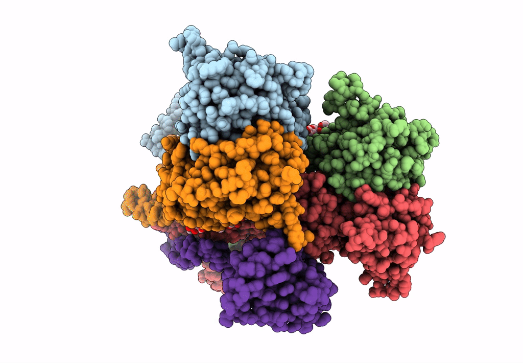
PDB ID:
7F5B
Keywords:
Title:
LBD-TMD focused reconstruction of DNQX-bound GluK2-1xNeto2 complex
Biological Source:
Source Organism(s):
Rattus norvegicus (Taxon ID: 10116)
Expression System(s):
Method Details:
Experimental Method:
Resolution:
3.90 Å
Aggregation State:
PARTICLE
Reconstruction Method:
SINGLE PARTICLE