7F2R image
Deposition Date 2021-06-14
Release Date 2021-09-01
Last Version Date 2024-11-06
Entry Detail
PDB ID:
7F2R
Keywords:
Title:
Crystal structure of VinK-VinL covalent complex formed with a pantetheineamide cross-linking probe
Biological Source:
Source Organism:
Method Details:
Experimental Method:
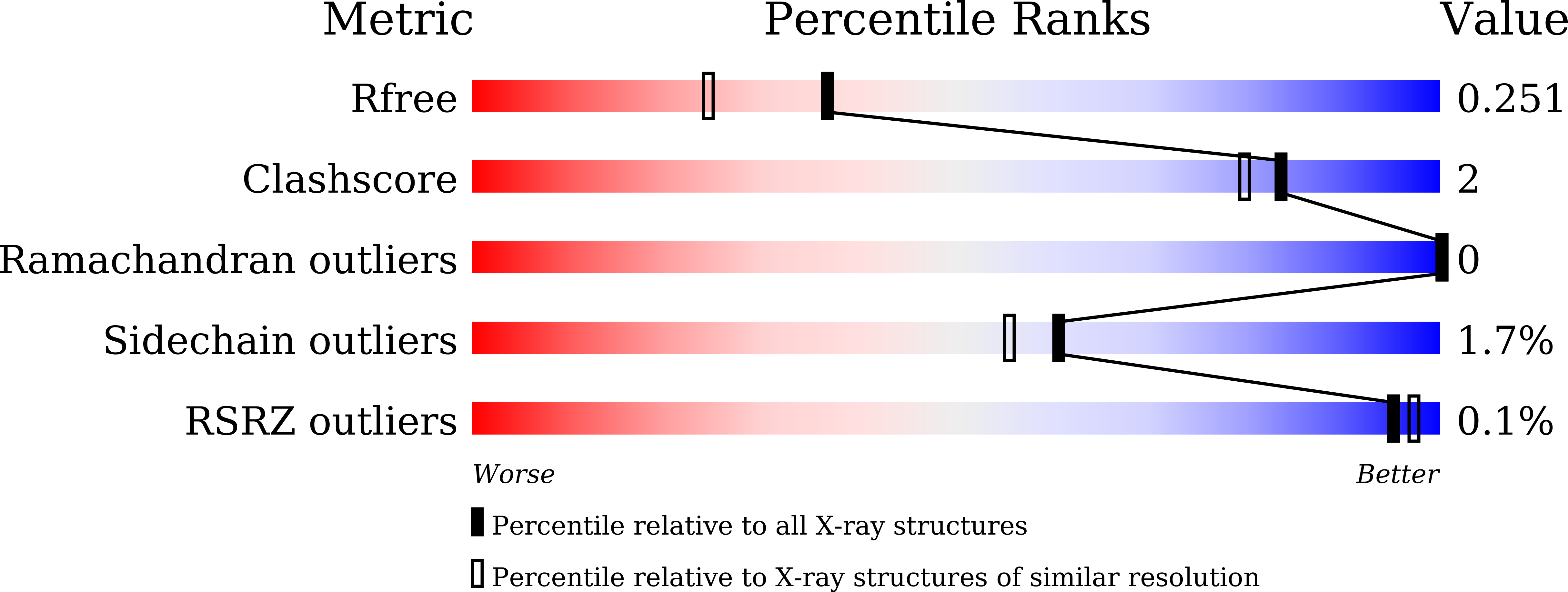
Resolution:
1.95 Å
R-Value Free:
0.24
R-Value Work:
0.19
R-Value Observed:
0.19
Space Group:
P 1 21 1
Macromolecular Entities
Structures with similar UniProt ID
Protein Blast
Polymer Type:polypeptide(L)
Molecule:Malonyl-CoA-[acyl-carrier-protein] transacylase
Gene (Uniprot):vinK
Mutagens:S106C
Chain IDs:A, C
Chain Length:328
Number of Molecules:2
Biological Source:Streptomyces halstedii
Structures with similar UniProt ID
Protein Blast
Polymer Type:polypeptide(L)
Molecule:Acyl-carrier-protein
Gene (Uniprot):vinL
Chain IDs:B, D
Chain Length:85
Number of Molecules:2
Biological Source:Streptomyces halstedii
Ligand Molecules
Primary Citation
Complex structure of the acyltransferase VinK and the carrier protein VinL with a pantetheine cross-linking probe.
Acta Crystallogr.,Sect.F 77 294 302 (2021)
PMID: 34473106 DOI: 10.1107/S2053230X21008761

Abstact

Acyltransferases are responsible for the selection and loading of acyl units onto carrier proteins in polyketide and fatty-acid biosynthesis. Despite the importance of protein-protein interactions between the acyltransferase and the carrier protein, structural information on acyltransferase-carrier protein interactions is limited because of the transient interactions between them. In the biosynthesis of the polyketide vicenistatin, the acyltransferase VinK recognizes the carrier protein VinL for the transfer of a dipeptidyl unit. The crystal structure of a VinK-VinL covalent complex formed with a 1,2-bismaleimidoethane cross-linking reagent has been determined previously. Here, the crystal structure of a VinK-VinL covalent complex formed with a pantetheine cross-linking probe is reported at 1.95 Å resolution. In the structure of the VinK-VinL-probe complex, the pantetheine probe that is attached to VinL is covalently connected to the side chain of the mutated Cys106 of VinK. The interaction interface between VinK and VinL is essentially the same in the two VinK-VinL complex structures, although the position of the pantetheine linker slightly differs. This structural observation suggests that interface interactions are not affected by the cross-linking strategy used.

Legend

Protein

Chemical

Disease

Primary Citation of related structures
Feedback Form
Name
Email
Institute
Feedback