Deposition Date
2021-06-11
Release Date
2022-04-20
Last Version Date
2024-10-16
Entry Detail
PDB ID:
7F2G
Keywords:
Title:
Crystal structure of the sensor domain of VbrK from Vibrio rotiferianus (crystal type 1)
Biological Source:
Source Organism(s):
Vibrio rotiferianus (Taxon ID: 190895)
Expression System(s):
Method Details:
Experimental Method:
Resolution:
1.90 Å
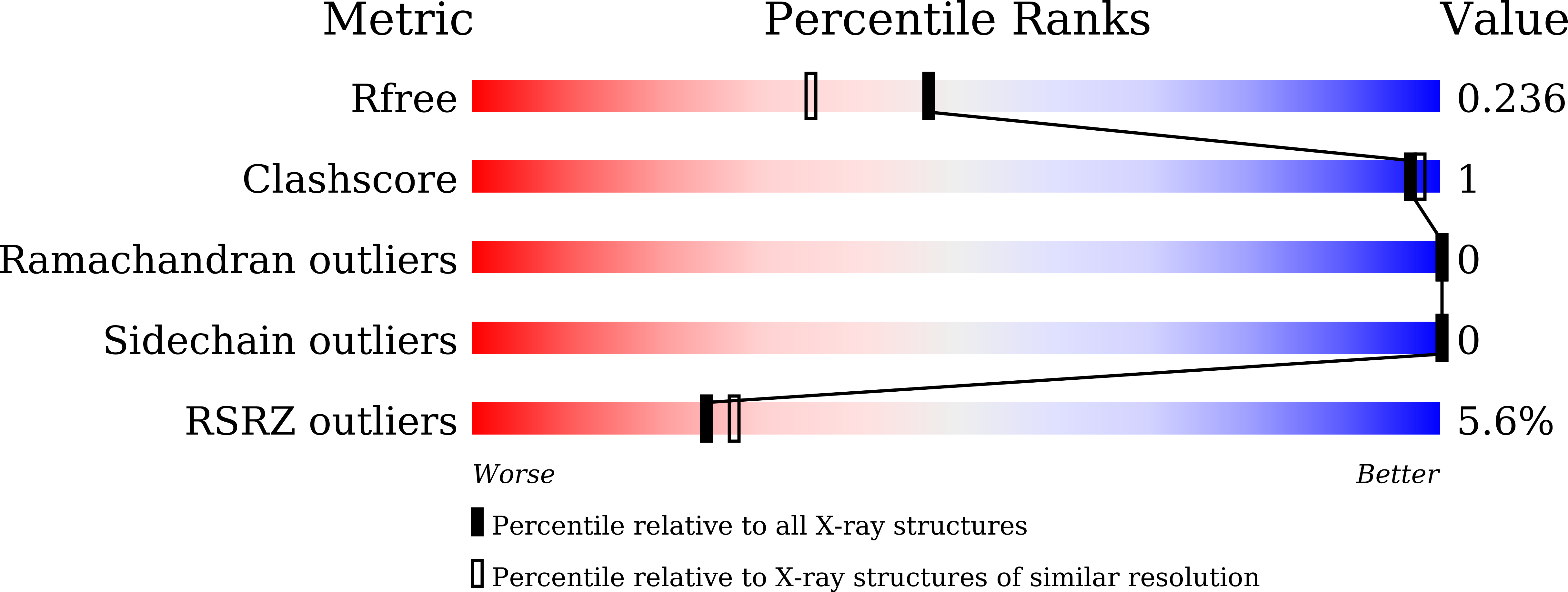
R-Value Free:
0.23
R-Value Work:
0.19
R-Value Observed:
0.19
Space Group:
P 21 21 21