Deposition Date
2021-06-10
Release Date
2022-06-15
Last Version Date
2025-07-02
Entry Detail
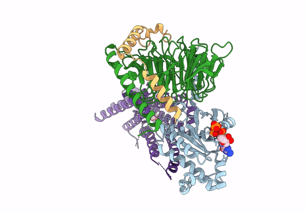
PDB ID:
7F24
Keywords:
Title:
Cryo-EM structure of the GTP-bound dopamine receptor 1 and mini-Gs complex without Nb35
Biological Source:
Source Organism(s):
Homo sapiens (Taxon ID: 9606)
Expression System(s):
Method Details:
Experimental Method:
Resolution:
4.16 Å
Aggregation State:
PARTICLE
Reconstruction Method:
SINGLE PARTICLE