Deposition Date
2021-06-09
Release Date
2022-04-20
Last Version Date
2025-09-17
Entry Detail
PDB ID:
7F1V
Keywords:
Title:
Crystal structure of Pseudomonas putida methionine gamma-lyase Q349S mutant with L-homocysteine intermediates
Biological Source:
Source Organism(s):
Pseudomonas putida (Taxon ID: 303)
Expression System(s):
Method Details:
Experimental Method:
Resolution:
2.25 Å
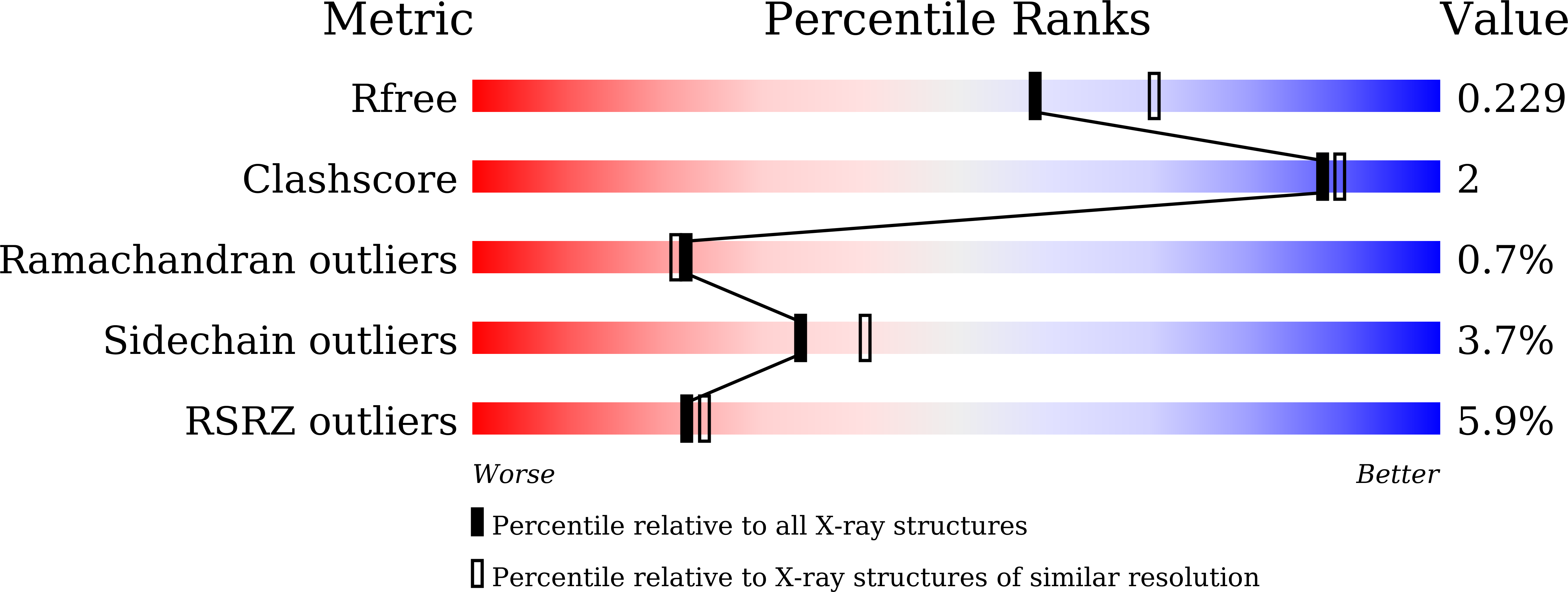
R-Value Free:
0.22
R-Value Work:
0.18
R-Value Observed:
0.18
Space Group:
P 21 21 2