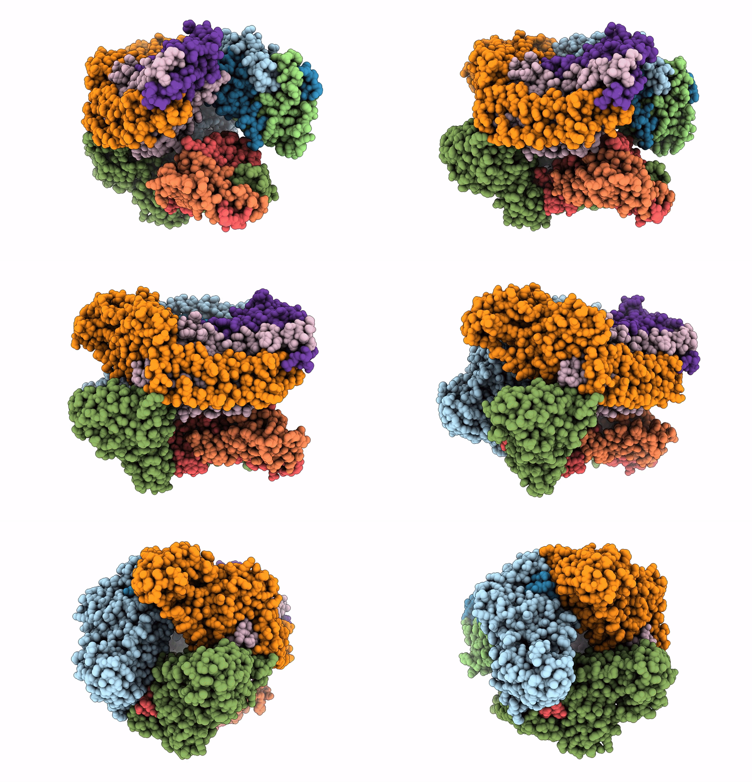
7EV9 image
Deposition Date 2021-05-20
Release Date 2021-07-21
Last Version Date 2024-06-05
Entry Detail
PDB ID:
7EV9
Keywords:
Title:
cryoEM structure of particulate methane monooxygenase associated with Cu(I)
Biological Source:
Method Details:
Experimental Method:
Resolution:
2.60 Å
Aggregation State:
PARTICLE
Reconstruction Method:
SINGLE PARTICLE
Macromolecular Entities
Polymer Type:polypeptide(L)
Molecule:Particulate methane monooxygenase alpha subunit
Gene (Uniprot):pmoB1, pmoB2
Chain IDs:A, D (auth: E), G (auth: I)
Chain Length:414
Number of Molecules:3
Biological Source:Methylococcus capsulatus (strain ATCC 33009 / NCIMB 11132 / Bath)
Polymer Type:polypeptide(L)
Molecule:Particulate methane monooxygenase beta subunit
Gene (Uniprot):pmoA1, pmoA2
Chain IDs:B, E (auth: F), H (auth: J)
Chain Length:247
Number of Molecules:3
Biological Source:Methylococcus capsulatus (strain ATCC 33009 / NCIMB 11132 / Bath)
Polymer Type:polypeptide(L)
Molecule:Ammonia monooxygenase/methane monooxygenase, subunit C family protein
Gene (Uniprot):pmoC1
Chain IDs:C, F (auth: G), I (auth: K)
Chain Length:260
Number of Molecules:3
Biological Source:Methylococcus capsulatus (strain ATCC 33009 / NCIMB 11132 / Bath)
Ligand Molecules
Primary Citation
Copper Centers in the Cryo-EM Structure of Particulate Methane Monooxygenase Reveal the Catalytic Machinery of Methane Oxidation.
J.Am.Chem.Soc. 143 9922 9932 (2021)
PMID: 34170126 DOI: 10.1021/jacs.1c04082

Abstact

The particulate methane monooxygenase (pMMO) is the first enzyme in the C1 metabolic pathway in methanotrophic bacteria. As this enzyme converts methane into methanol efficiently near room temperature, it has become the paradigm for developing an understanding of this difficult C1 chemistry. pMMO is a membrane-bound protein with three subunits (PmoB, PmoA, and PmoC) and 12-14 coppers distributed among different sites. X-ray crystal structures that have revealed only three mononuclear coppers at three sites have neither disclosed the location of the active site nor the catalytic mechanism of the enzyme. Here we report a cyro-EM structure of holo-pMMO from Methylococcus capsulatus (Bath) at 2.5 Å, and develop quantitative electrostatic-potential profiling to scrutinize the nonprotein densities for signatures of the copper cofactors. Our results confirm a mononuclear CuI at the A site, resolve two CuIs at the B site, and uncover additional CuI clusters at the PmoA/PmoC interface within the membrane (D site) and in the water-exposed C-terminal subdomain of the PmoB (E clusters). These findings complete the minimal set of copper factors required for catalytic turnover of pMMO, offering a glimpse of the catalytic machinery for methane oxidation according to the chemical principles underlying the mechanism proposed earlier.

Legend

Protein

Chemical

Disease

Primary Citation of related structures