7ERV image
Entry Detail
PDB ID:
7ERV
Keywords:
Title:
Crystal structure of L-histidine decarboxylase (C57S/C101V/C282V mutant) from Photobacterium phosphoreum
Biological Source:
Host Organism:
PDB Version:
Deposition Date:
2021-05-07
Release Date:
2022-02-16
Method Details:
Experimental Method:
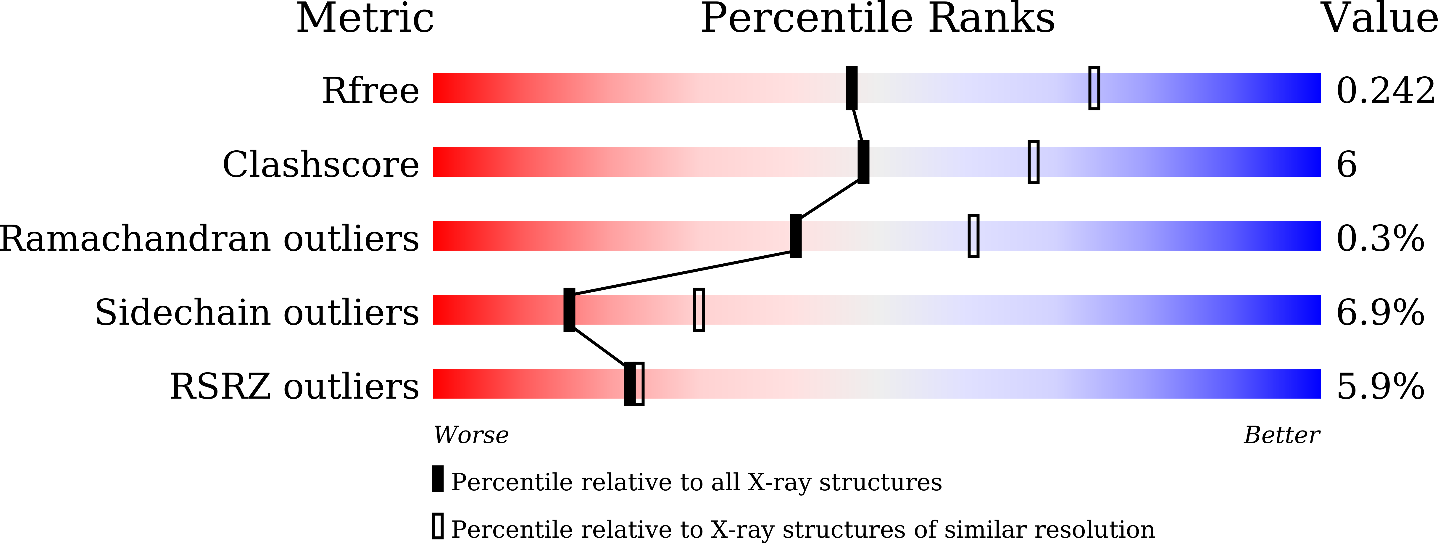
Resolution:
2.50 Å
R-Value Free:
0.23
R-Value Work:
0.18
R-Value Observed:
0.18
Space Group:
P 61 2 2
Macromolecular Entities
Polymer Type:polypeptide(L)
Description:Histidine decarboxylase
Mutations:C57S/C101V/C282V
Chain IDs:A
Chain Length:374
Number of Molecules:1
Biological Source:Photobacterium phosphoreum
Modified Residue
Compound ID Chain ID Parent Comp ID Details 2D Image
LLP A LYS modified residue
Ligand Molecules
Primary Citation
Structural insights into the enhanced thermostability of cysteine substitution mutants of L-histidine decarboxylase from Photobacterium phosphoreum.
J.Biochem. 171 31 40 (2022)
PMID: 34622278 DOI: 10.1093/jb/mvab103

Abstact

Enzymatic amino acid assays are important in physiological research and clinical diagnostics because abnormal amino acid concentrations in biofluids are associated with various diseases. L-histidine decarboxylase from Photobacterium phosphoreum (PpHDC) is a pyridoxal 5'-phosphate-dependent enzyme and a candidate for use in an L-histidine quantitation assay. Previous cysteine substitution experiments demonstrated that the PpHDC C57S mutant displayed improved long-term storage stability and thermostability when compared with those of the wild-type enzyme. In this study, combinational mutation experiments of single cysteine substitution mutants of PpHDC were performed, revealing that the PpHDC C57S/C101V/C282V mutant possessed the highest thermostability. The stabilizing mechanism of these mutations was elucidated by solving the structures of PpHDC C57S and C57S/C101V/C282V mutants by X-ray crystallography. In the crystal structures, two symmetry-related PpHDC molecules form a domain-swapped homodimer. The side chain of S57 is solvent exposed in the structure, indicating that the C57S mutation eliminates chemical oxidation or disulfide bond formation with a free thiol group, thereby providing greater stability. Residues 101 and 282 form hydrophobic interactions with neighboring hydrophobic residues. Mutations C101V and C282V enhanced thermostability of PpHDC by filling a cavity present in the hydrophobic core (C101V) and increasing hydrophobic interactions.

Legend

Protein

Chemical

Disease

Primary Citation of related structures