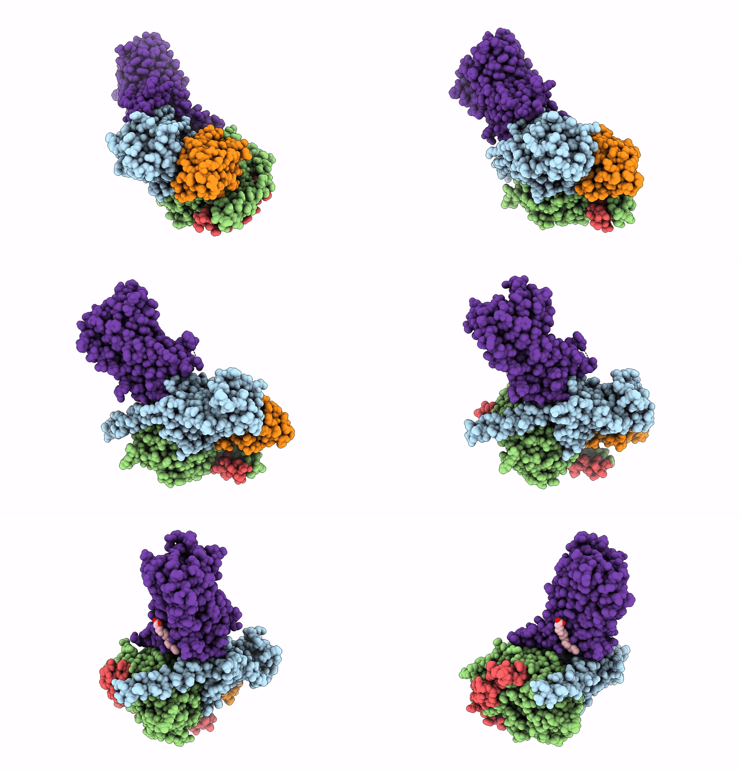
7EPT image
Deposition Date 2021-04-27
Release Date 2022-05-11
Last Version Date 2025-07-02
Entry Detail
PDB ID:
7EPT
Title:
Structural basis for the tethered peptide activation of adhesion GPCRs
Biological Source:
Source Organism:
Host Organism:
Method Details:
Experimental Method:
Resolution:
3.00 Å
Aggregation State:
PARTICLE
Reconstruction Method:
SINGLE PARTICLE
Macromolecular Entities
Polymer Type:polypeptide(L)
Molecule:Guanine nucleotide-binding protein G(s) subunit alpha isoforms short
Chain IDs:A
Chain Length:394
Number of Molecules:1
Biological Source:Homo sapiens
Polymer Type:polypeptide(L)
Molecule:Guanine nucleotide-binding protein G(I)/G(S)/G(T) subunit beta-1
Gene (Uniprot):GNB1
Chain IDs:B
Chain Length:358
Number of Molecules:1
Biological Source:Homo sapiens
Polymer Type:polypeptide(L)
Molecule:Guanine nucleotide-binding protein G(I)/G(S)/G(O) subunit gamma-2
Gene (Uniprot):GNG2
Chain IDs:C (auth: G)
Chain Length:71
Number of Molecules:1
Biological Source:Homo sapiens
Polymer Type:polypeptide(L)
Molecule:Nanobody-35
Chain IDs:D (auth: N)
Chain Length:128
Number of Molecules:1
Biological Source:synthetic construct
Polymer Type:polypeptide(L)
Molecule:Adhesion G-protein coupled receptor D1
Gene (Uniprot):ADGRD1
Chain IDs:E (auth: R)
Chain Length:283
Number of Molecules:1
Biological Source:Homo sapiens
Ligand Molecules
Primary Citation
Structural basis for the tethered peptide activation of adhesion GPCRs.
Nature 604 763 770 (2022)
PMID: 35418678 DOI: 10.1038/s41586-022-04619-y

Abstact

Adhesion G-protein-coupled receptors (aGPCRs) are important for organogenesis, neurodevelopment, reproduction and other processes1-6. Many aGPCRs are activated by a conserved internal (tethered) agonist sequence known as the Stachel sequence7-12. Here, we report the cryogenic electron microscopy (cryo-EM) structures of two aGPCRs in complex with Gs: GPR133 and GPR114. The structures indicate that the Stachel sequences of both receptors assume an α-helical-bulge-β-sheet structure and insert into a binding site formed by the transmembrane domain (TMD). A hydrophobic interaction motif (HIM) within the Stachel sequence mediates most of the intramolecular interactions with the TMD. Combined with the cryo-EM structures, biochemical characterization of the HIM motif provides insight into the cross-reactivity and selectivity of the Stachel sequences. Two interconnected mechanisms, the sensing of Stachel sequences by the conserved 'toggle switch' W6.53 and the constitution of a hydrogen-bond network formed by Q7.49/Y7.49 and the P6.47/V6.47φφG6.50 motif (φ indicates a hydrophobic residue), are important in Stachel sequence-mediated receptor activation and Gs coupling. Notably, this network stabilizes kink formation in TM helices 6 and 7 (TM6 and TM7, respectively). A common Gs-binding interface is observed between the two aGPCRs, and GPR114 has an extended TM7 that forms unique interactions with Gs. Our structures reveal the detailed mechanisms of aGPCR activation by Stachel sequences and their Gs coupling.

Legend

Protein

Chemical

Disease

Primary Citation of related structures