Deposition Date
1990-11-13
Release Date
1992-04-15
Last Version Date
2024-03-06
Entry Detail
PDB ID:
7ENL
Keywords:
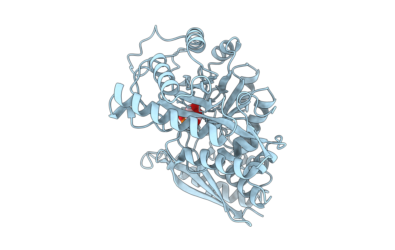
Title:
MECHANISM OF ENOLASE: THE CRYSTAL STRUCTURE OF ENOLASE-MG2+-PHOSPHOGLYCERATE(SLASH) PHOSPHOENOLPYRUVATE COMPLEX AT 2.2-ANGSTROMS RESOLUTION
Biological Source:
Source Organism(s):
Saccharomyces cerevisiae (Taxon ID: 4932)
Method Details:
Experimental Method:
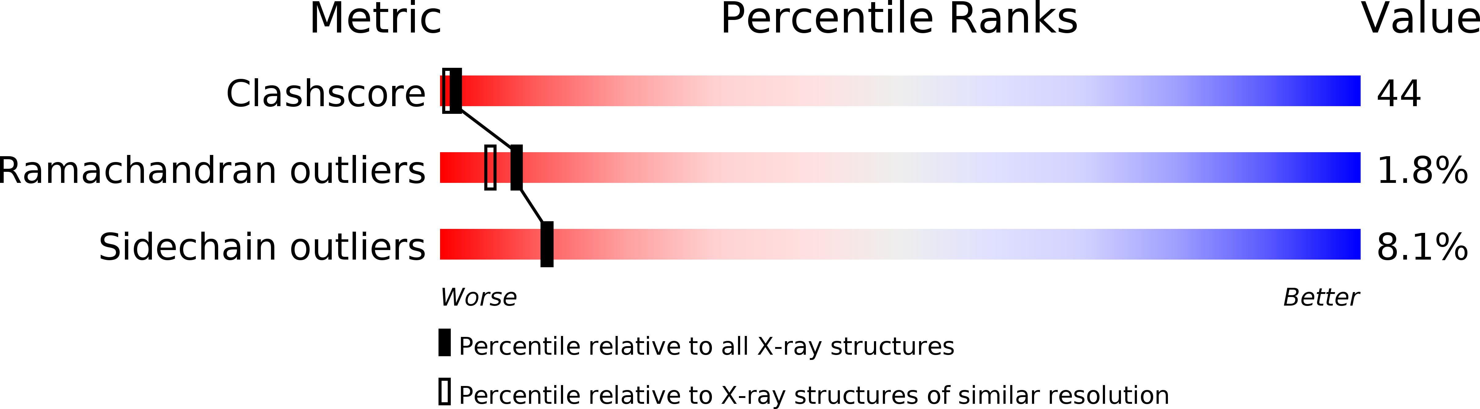
Resolution:
2.20 Å
R-Value Observed:
0.16
Space Group:
P 42 21 2