7EFQ image
Entry Detail
PDB ID:
7EFQ
Keywords:
Title:
Crystal structure of hPPARgamma ligand binding domain complexed with rosiglitazone-based fluorescence probe
Biological Source:
Source Organism:
Host Organism:
PDB Version:
Deposition Date:
2021-03-23
Release Date:
2021-05-12
Method Details:
Experimental Method:
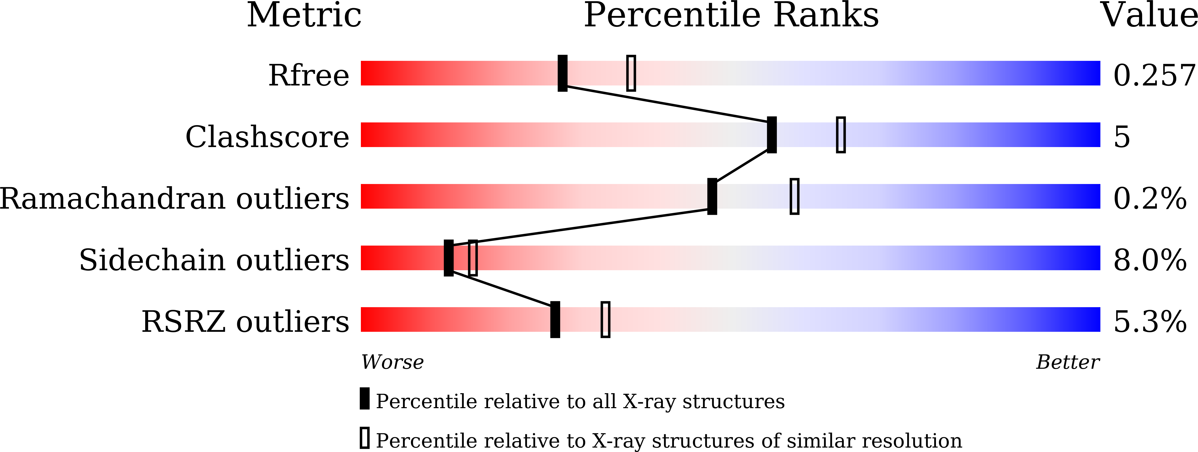
Resolution:
2.30 Å
R-Value Free:
0.26
R-Value Work:
0.22
Space Group:
C 1 2 1
Macromolecular Entities
Polymer Type:polypeptide(L)
Description:Peroxisome proliferator-activated receptor gamma
Chain IDs:A, B
Chain Length:276
Number of Molecules:2
Biological Source:Homo sapiens
Ligand Molecules
Primary Citation
Synthesis of a Coumarin-Based PPAR gamma Fluorescence Probe for Competitive Binding Assay.
Int J Mol Sci 22 ? ? (2021)
PMID: 33919837 DOI: 10.3390/ijms22084034

Abstact

Peroxisome proliferator-activated receptor γ (PPARγ) is a molecular target of metabolic syndrome and inflammatory disease. PPARγ is an important nuclear receptor and numerous PPARγ ligands were developed to date; thus, efficient assay methods are important. Here, we investigated the incorporation of 7-diethylamino coumarin into the PPARγ agonist rosiglitazone and used the compound in a binding assay for PPARγ. PPARγ-ligand-incorporated 7-methoxycoumarin, 1, showed weak fluorescence intensity in a previous report. We synthesized PPARγ-ligand-incorporating coumarin, 2, in this report, and it enhanced the fluorescence intensity. The PPARγ ligand 2 maintained the rosiglitazone activity. The obtained partial agonist 6 appeared to act through a novel mechanism. The fluorescence intensity of 2 and 6 increased by binding to the ligand binding domain (LBD) of PPARγ and the affinity of reported PPARγ ligands were evaluated using the probe.

Legend

Protein

Chemical

Disease

Primary Citation of related structures