7EFA image
Deposition Date 2021-03-21
Release Date 2021-06-23
Last Version Date 2023-11-29
Entry Detail
PDB ID:
7EFA
Title:
Crystal structure of the complex between the C-terminal domain of mouse MUTYH and human PCNA
Biological Source:
Source Organism:
Homo sapiens (Taxon ID: 9606)
Mus musculus (Taxon ID: 10090)
Host Organism:
Method Details:
Experimental Method:
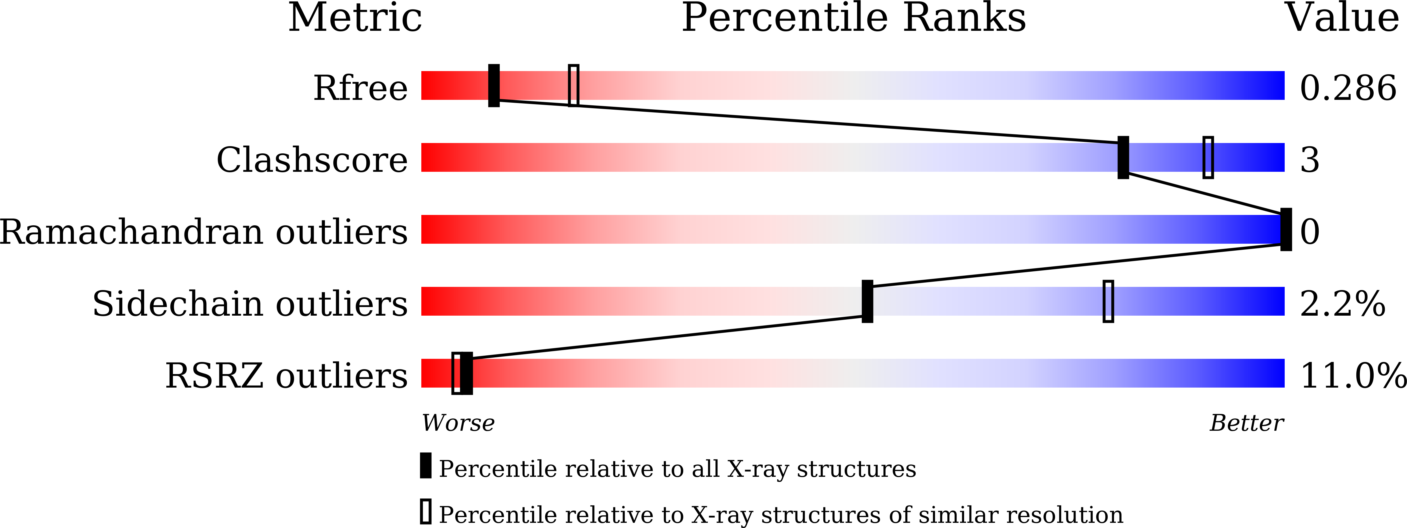
Resolution:
2.70 Å
R-Value Free:
0.28
R-Value Work:
0.23
R-Value Observed:
0.23
Space Group:
P 63
Macromolecular Entities
Polymer Type:polypeptide(L)
Molecule:Proliferating cell nuclear antigen
Gene (Uniprot):PCNA
Chain IDs:A
Chain Length:261
Number of Molecules:1
Biological Source:Homo sapiens
Polymer Type:polypeptide(L)
Molecule:Adenine DNA glycosylase
Gene (Uniprot):Mutyh
Chain IDs:B
Chain Length:189
Number of Molecules:1
Biological Source:Mus musculus
Primary Citation
Structure of the mammalian adenine DNA glycosylase MUTYH: insights into the base excision repair pathway and cancer.
Nucleic Acids Res. 49 7154 7163 (2021)
PMID: 34142156 DOI: 10.1093/nar/gkab492

Abstact

Mammalian MutY homologue (MUTYH) is an adenine DNA glycosylase that excises adenine inserted opposite 8-oxoguanine (8-oxoG). The inherited variations in human MUTYH gene are known to cause MUTYH-associated polyposis (MAP), which is associated with colorectal cancer. MUTYH is involved in base excision repair (BER) with proliferating cell nuclear antigen (PCNA) in DNA replication, which is unique and critical for effective mutation-avoidance. It is also reported that MUTYH has a Zn-binding motif in a unique interdomain connector (IDC) region, which interacts with Rad9-Rad1-Hus1 complex (9-1-1) in DNA damage response, and with apurinic/apyrimidinic endonuclease 1 (APE1) in BER. However, the structural basis for the BER pathway by MUTYH and its interacting proteins is unclear. Here, we determined the crystal structures of complexes between mouse MUTYH and DNA, and between the C-terminal domain of mouse MUTYH and human PCNA. The structures elucidated the repair mechanism for the A:8-oxoG mispair including DNA replication-coupled repair process involving MUTYH and PCNA. The Zn-binding motif was revealed to comprise one histidine and three cysteine residues. The IDC, including the Zn-binding motif, is exposed on the MUTYH surface, suggesting its interaction modes with 9-1-1 and APE1, respectively. The structure of MUTYH explains how MAP mutations perturb MUTYH function.

Legend

Protein

Chemical

Disease

Primary Citation of related structures