Deposition Date
2021-03-19
Release Date
2022-03-23
Last Version Date
2024-05-29
Entry Detail
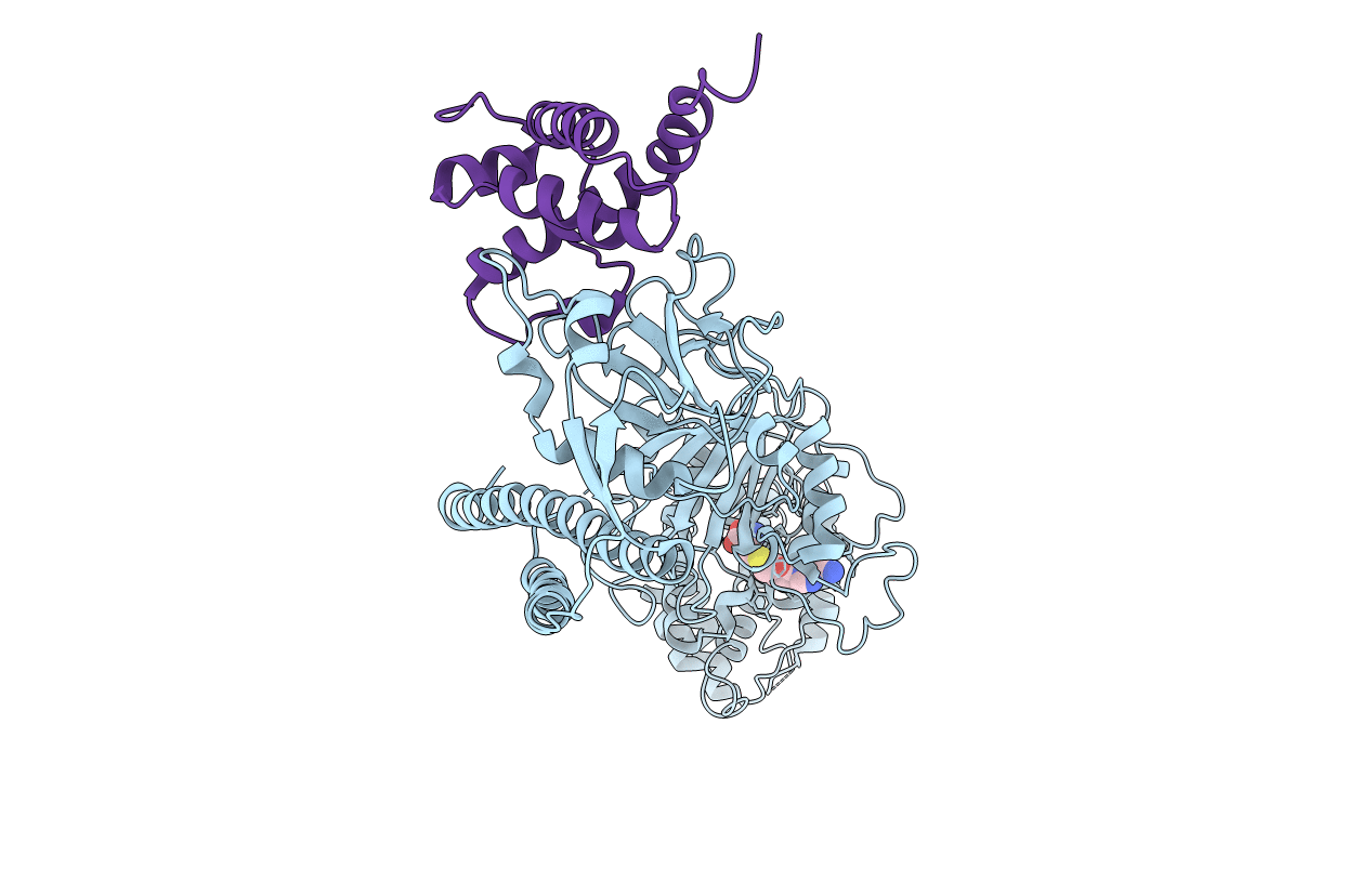
PDB ID:
7EEW
Keywords:
Title:
Crystal structure of the intact MTase from Vibrio vulnificus YJ016 in complex with the DNA-mimicking Ocr protein and the S-adenosyl-L-homocysteine (SAH)
Biological Source:
Source Organism(s):
Vibrio vulnificus (strain YJ016) (Taxon ID: 196600)
Escherichia phage T7 (Taxon ID: 10760)
Escherichia phage T7 (Taxon ID: 10760)
Expression System(s):
Method Details:
Experimental Method:
Resolution:
2.90 Å
R-Value Free:
0.29
R-Value Work:
0.27
R-Value Observed:
0.27
Space Group:
P 65 2 2