Deposition Date
2021-03-09
Release Date
2021-07-21
Last Version Date
2023-11-29
Entry Detail
PDB ID:
7EB9
Keywords:
Title:
The structure of the A20-binding inhibitor of NF-kB 1 in complex with tetra-ubiquitin
Biological Source:
Source Organism(s):
Homo sapiens (Taxon ID: 9606)
Expression System(s):
Method Details:
Experimental Method:
Resolution:
3.20 Å
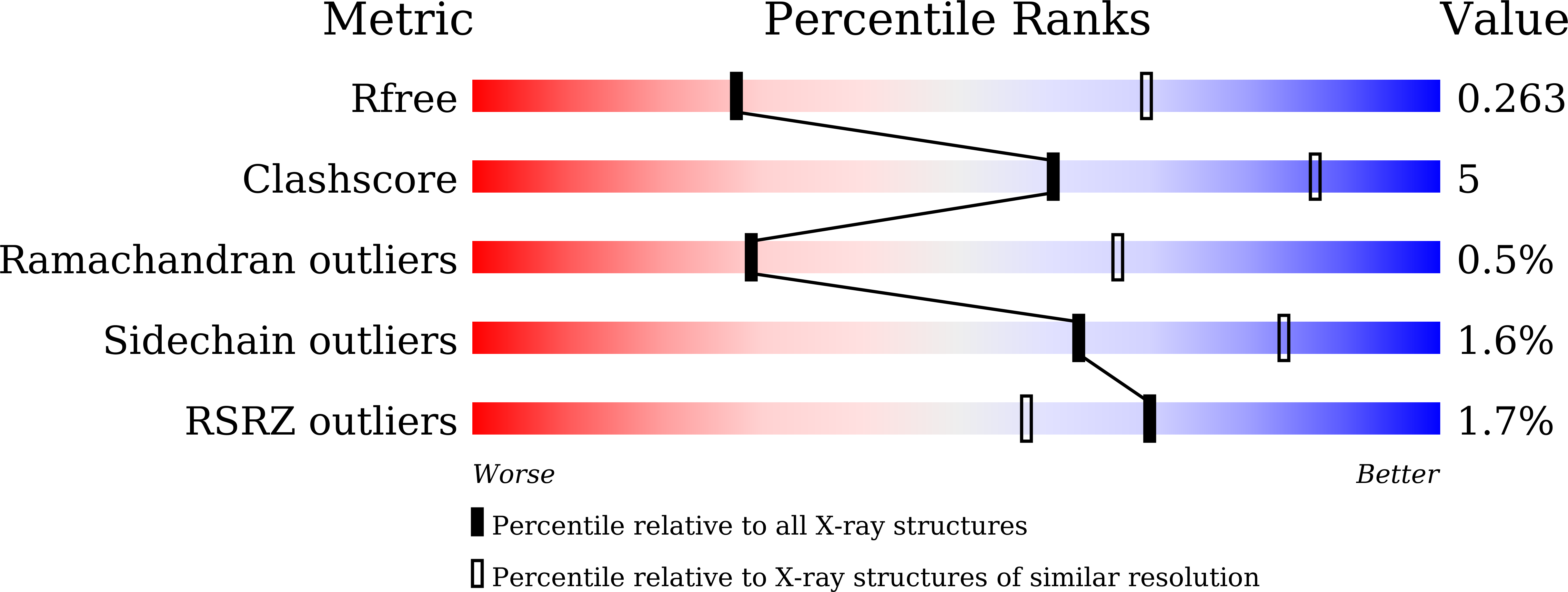
R-Value Free:
0.26
R-Value Work:
0.21
R-Value Observed:
0.22
Space Group:
P 2 21 21