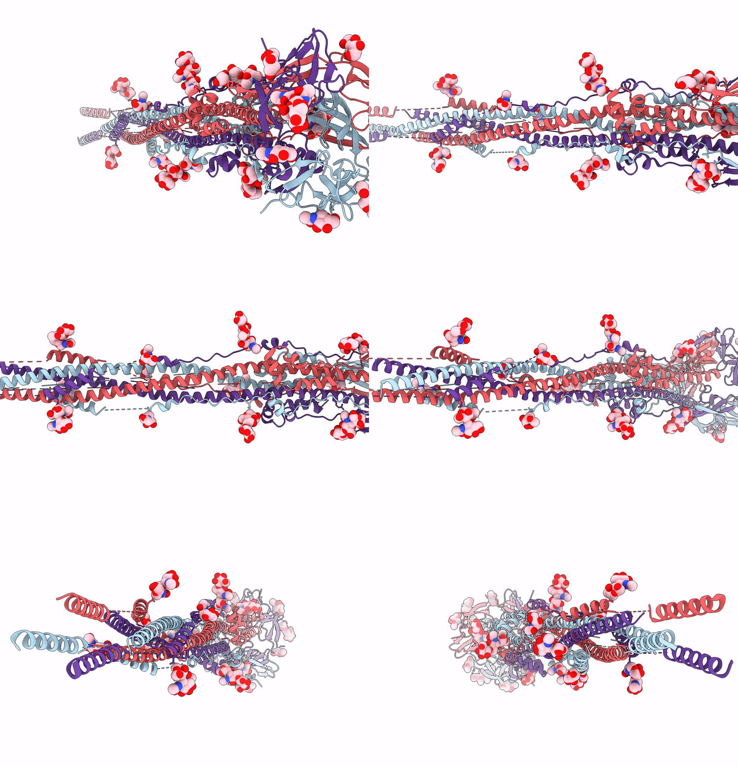
7E9T image
Deposition Date 2021-03-05
Release Date 2021-11-17
Last Version Date 2024-10-16
Entry Detail
PDB ID:
7E9T
Keywords:
Title:
Nanometer resolution in situ structure of SARS-CoV-2 post-fusion spike
Biological Source:
Method Details:
Experimental Method:
Resolution:
10.90 Å
Aggregation State:
PARTICLE
Reconstruction Method:
SUBTOMOGRAM AVERAGING
Macromolecular Entities
Polymer Type:polypeptide(L)
Molecule:Spike protein S2
Gene (Uniprot):S
Chain IDs:A, B, C
Chain Length:532
Number of Molecules:3
Biological Source:Severe acute respiratory syndrome coronavirus 2
Ligand Molecules
Primary Citation
Nanometer-resolution in situ structure of the SARS-CoV-2 postfusion spike protein.
Proc.Natl.Acad.Sci.USA 118 ? ? (2021)
PMID: 34782481 DOI: 10.1073/pnas.2112703118

Abstact

The spike protein of severe acute respiratory syndrome coronavirus 2 (SARS-CoV-2) mediates membrane fusion to allow entry of the viral genome into host cells. To understand its detailed entry mechanism and develop a specific entry inhibitor, in situ structural information on the SARS-CoV-2 spike protein in different states is urgent. Here, by using cryo-electron tomography, we observed both prefusion and postfusion spikes in β-propiolactone-inactivated SARS-CoV-2 virions and solved the in situ structure of the postfusion spike at nanometer resolution. Compared to previous reports, the six-helix bundle fusion core, the glycosylation sites, and the location of the transmembrane domain were clearly resolved. We observed oligomerization patterns of the spikes on the viral membrane, likely suggesting a mechanism of fusion pore formation.

Legend

Protein

Chemical

Disease

Primary Citation of related structures