
Deposition Date
2021-03-04
Release Date
2021-06-23
Last Version Date
2024-10-23
Entry Detail
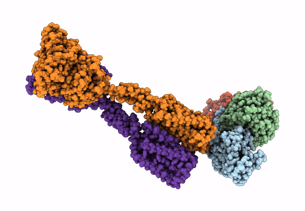
PDB ID:
7E9H
Keywords:
Title:
Cryo-EM structure of Gi-bound metabotropic glutamate receptor mGlu4
Biological Source:
Source Organism(s):
Homo sapiens (Taxon ID: 9606)
Expression System(s):
Method Details:
Experimental Method:
Resolution:
4.00 Å
Aggregation State:
PARTICLE
Reconstruction Method:
SINGLE PARTICLE
