Deposition Date
2021-02-15
Release Date
2021-07-07
Last Version Date
2023-11-29
Entry Detail
PDB ID:
7E4R
Keywords:
Title:
Crystal structure of tubulin in complex with D-DM1-SMe
Biological Source:
Source Organism(s):
Bos taurus (Taxon ID: 9913)
Rattus norvegicus (Taxon ID: 10116)
Gallus gallus (Taxon ID: 9031)
Rattus norvegicus (Taxon ID: 10116)
Gallus gallus (Taxon ID: 9031)
Expression System(s):
Method Details:
Experimental Method:
Resolution:
2.60 Å
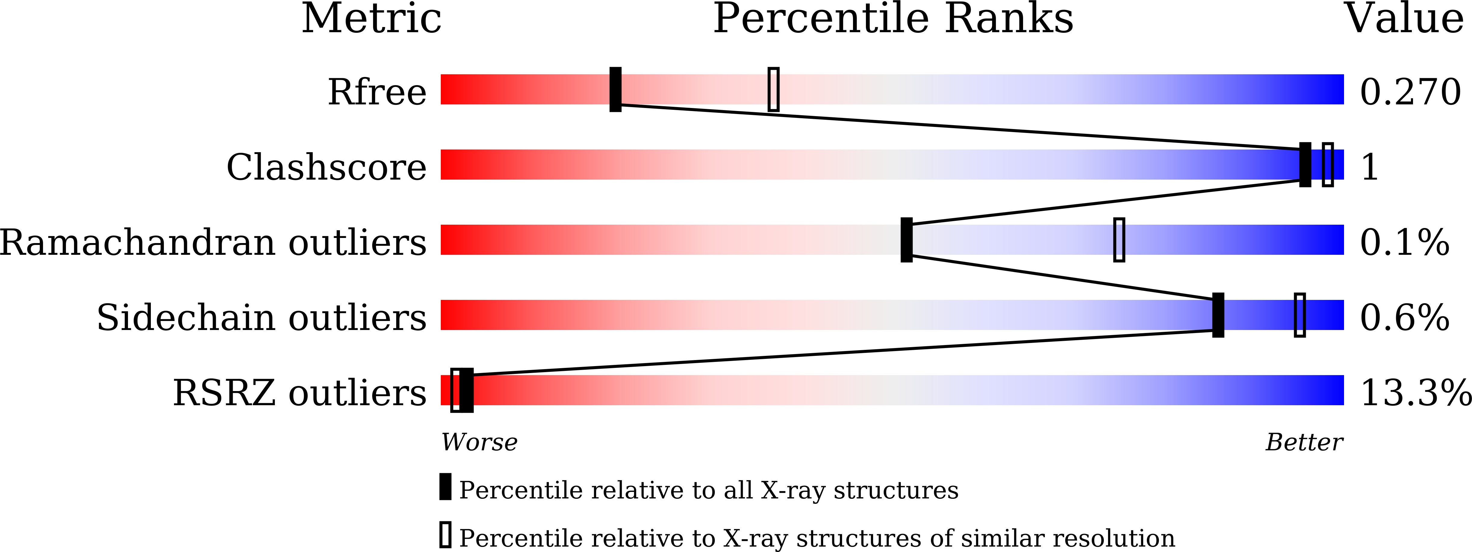
R-Value Free:
0.26
R-Value Work:
0.22
R-Value Observed:
0.22
Space Group:
P 21 21 21