
Deposition Date
2021-02-09
Release Date
2022-02-16
Last Version Date
2023-11-29
Entry Detail
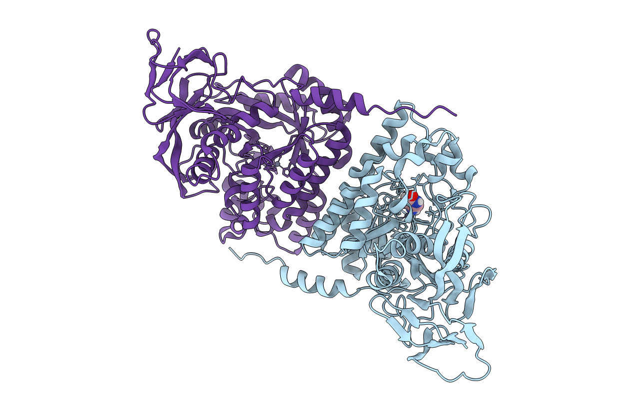
PDB ID:
7E3U
Keywords:
Title:
Crystal structure of the Pseudomonas aeruginosa dihydropyrimidinase complexed with 5-AU
Biological Source:
Source Organism(s):
Expression System(s):
Method Details:
Experimental Method:
Resolution:
2.16 Å
R-Value Free:
0.23
R-Value Work:
0.18
R-Value Observed:
0.18
Space Group:
P 31 2 1
