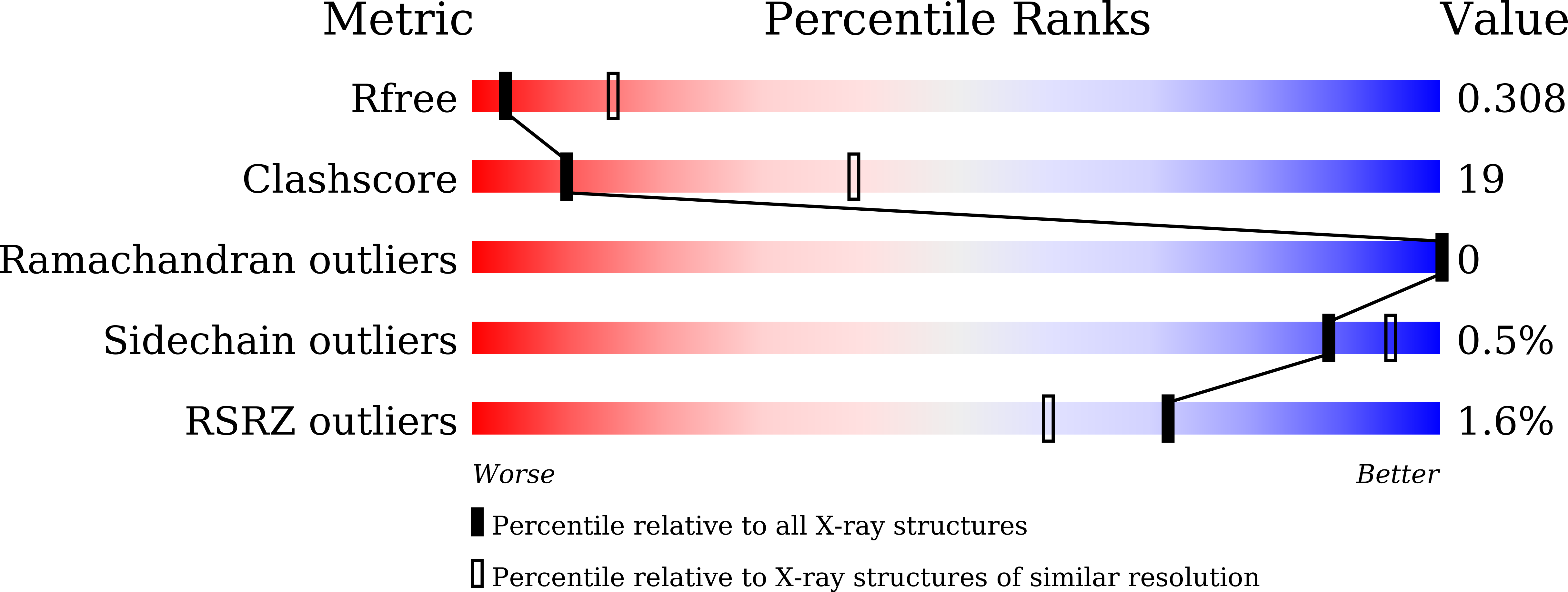
7E2O image
Entry Detail
PDB ID:
7E2O
Keywords:
Title:
X-ray Crystal structure of PPARgamma R288H mutant.
Biological Source:
Source Organism:
Host Organism:
PDB Version:
Deposition Date:
2021-02-06
Release Date:
2022-01-19
Method Details:
Experimental Method:
Resolution:
3.20 Å
R-Value Free:
0.31
R-Value Work:
0.23
R-Value Observed:
0.23
Space Group:
C 1 2 1
Macromolecular Entities
Polymer Type:polypeptide(L)
Description:Peroxisome proliferator-activated receptor gamma
Mutations:R288H
Chain IDs:A, B
Chain Length:276
Number of Molecules:2
Biological Source:Homo sapiens
Primary Citation
Structural Insights into the Loss-of-Function R288H Mutant of Human PPAR gamma.
Biol.Pharm.Bull. 44 1196 1201 (2021)
PMID: 34471047 DOI: 10.1248/bpb.b21-00253

Abstact

Peroxisome proliferator-activated receptor gamma (PPARγ) is a nuclear receptor and the molecular target of thiazolidinedione-class antidiabetic drugs. It has been reported that the loss of function R288H mutation in the human PPARγ ligand-binding domain (LBD) may be associated with the onset of colon cancer. A previous in vitro study showed that this mutation dampens 15-deoxy-Δ12,14-prostaglandin J2 (15d-PGJ2, a natural PPARγ agonist)-dependent transcriptional activation; however, it is poorly understood why the function of the R288H mutant is impaired and what role this arginine (Arg) residue plays. In this study, we found that the apo-form of R288H PPARγ mutant displays several altered conformational arrangements of the amino acid side chains in LBD: 1) the loss of a salt bridge between Arg288 and Glu295 leads to increased helix 3 movement; 2) closer proximity of Gln286 and His449 via a hydrogen bond, and closer proximity of Cys285 and Phe363 via hydrophobic interaction, stabilize the helix 3-helix 11 interaction; and 3) there is steric hindrance between Cys285/Gln286/Ser289/His449 and the flexible ligands 15d-PGJ2, 6-oxotetracosahexaenoic acid (6-oxoTHA), and 17-oxodocosahexaenoic acid (17-oxoDHA). These results suggest why Arg288 plays an important role in ligand binding and why the R288H mutation is disadvantageous for flexible ligand binding.

Legend

Protein

Chemical

Disease

Primary Citation of related structures