7E0F image
Entry Detail
PDB ID:
7E0F
EMDB ID:
Keywords:
Title:
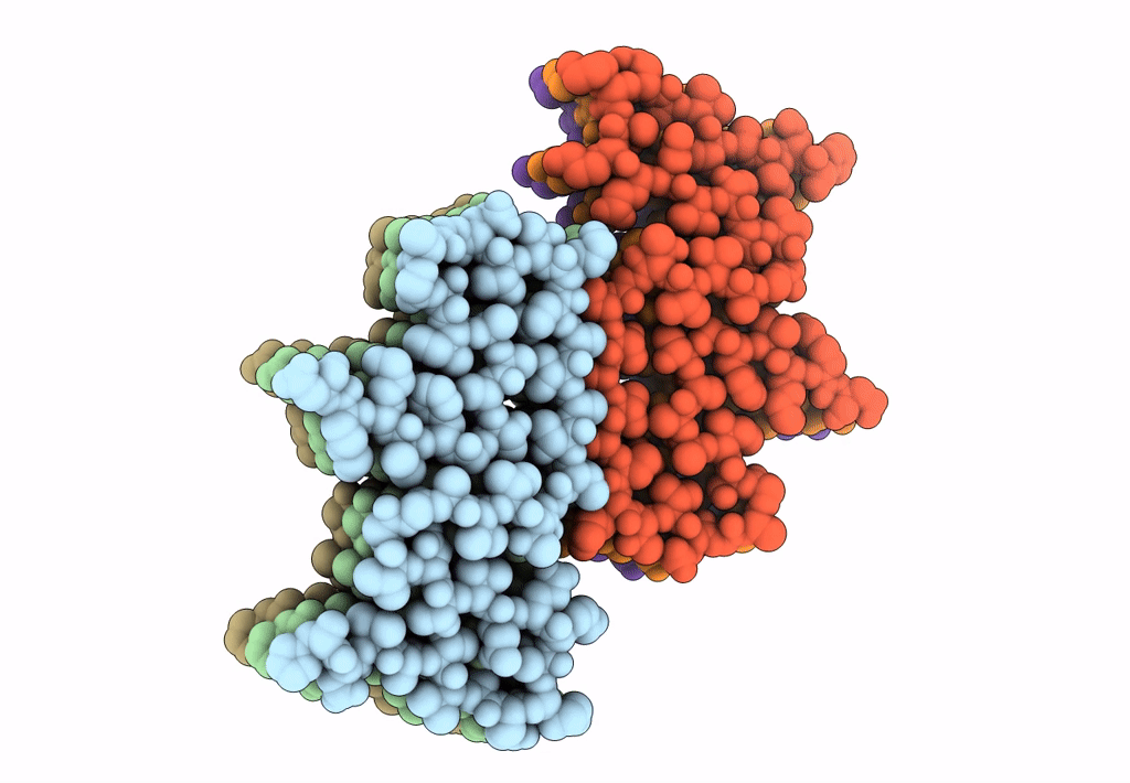
CryoEM structure of G51D alpha-synuclein amyloid fibril
Biological Source:
Source Organism:
Host Organism:
PDB Version:
Deposition Date:
2021-01-27
Release Date:
2021-10-06
Method Details:
Experimental Method:
Resolution:
3.02 Å
Aggregation State:
FILAMENT
Reconstruction Method:
HELICAL
Macromolecular Entities
Polymer Type:polypeptide(L)
Description:Alpha-synuclein
Mutations:G51D
Chain IDs:A, B, C, D, E, F
Chain Length:140
Number of Molecules:6
Biological Source:Homo sapiens
Ligand Molecules
Primary Citation
The hereditary mutation G51D unlocks a distinct fibril strain transmissible to wild-type alpha-synuclein.
Nat Commun 12 6252 6252 (2021)
PMID: 34716315 DOI: 10.1038/s41467-021-26433-2

Abstact

α-Synuclein (α-Syn) can form different fibril strains with distinct polymorphs and neuropathologies, which is associated with the clinicopathological variability in synucleinopathies. How different α-syn fibril strains are produced and selected under disease conditions remains poorly understood. In this study, we show that the hereditary mutation G51D induces α-syn to form a distinct fibril strain in vitro. The cryogenic electron microscopy (cryo-EM) structure of the G51D fibril strain was determined at 2.96 Å resolution. The G51D fibril displays a relatively small and extended serpentine fold distinct from other α-syn fibril structures. Moreover, we show by cryo-EM that wild-type (WT) α-syn can assembly into the G51D fibril strain via cross-seeding with G51D fibrils. Our study reveals a distinct structure of G51D fibril strain triggered by G51D mutation but feasibly adopted by both WT and G51D α-syn, which suggests the cross-seeding and strain selection of WT and mutant α-syn in familial Parkinson's disease (fPD).

Legend

Protein

Chemical

Disease

Primary Citation of related structures