7DWV image
Deposition Date 2021-01-18
Release Date 2021-10-13
Last Version Date 2024-11-13
Entry Detail
PDB ID:
7DWV
Keywords:
Title:
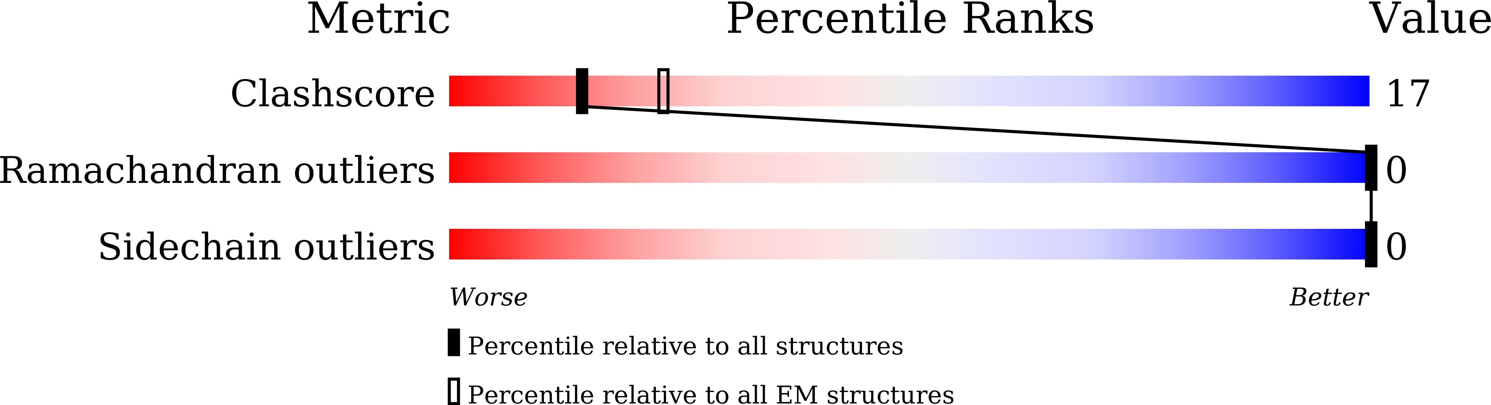
Cryo-EM structure of amyloid fibril formed by familial prion disease-related mutation E196K
Biological Source:
Source Organism:
Homo sapiens (Taxon ID: 9606)
Host Organism:
Method Details:
Experimental Method:
Resolution:
3.07 Å
Aggregation State:
HELICAL ARRAY
Reconstruction Method:
HELICAL
Macromolecular Entities
Structures with similar UniProt ID
Protein Blast
Polymer Type:polypeptide(L)
Molecule:Major prion protein
Gene (Uniprot):PRNP
Mutagens:E196K
Chain IDs:A, B, C, D, E, F
Chain Length:210
Number of Molecules:6
Biological Source:Homo sapiens
Ligand Molecules
Primary Citation
Genetic prion disease-related mutation E196K displays a novel amyloid fibril structure revealed by cryo-EM.
Sci Adv 7 eabg9676 eabg9676 (2021)
PMID: 34516876 DOI: 10.1126/sciadv.abg9676

Abstact

Prion diseases are caused by the conformational conversion of prion protein (PrP). Forty-two different mutations were identified in human PrP, leading to genetic prion diseases with distinct clinical syndromes. Here, we report the cryo–electron microscopy structure of an amyloid fibril formed by full-length human PrP with E196K mutation, a genetic Creutzfeldt-Jakob disease–related mutation. This mutation disrupts key interactions in the wild-type PrP fibril, forming an amyloid fibril with a conformation distinct from the wild-type PrP fibril and hamster brain–derived prion fibril. The E196K fibril consists of two protofibrils. Each subunit forms five β strands stabilized by a disulfide bond and an unusual hydrophilic cavity stabilized by a salt bridge. Four pairs of amino acids from opposing subunits form four salt bridges to stabilize the zigzag interface of the two protofibrils. Our results provide structural evidences of the diverse prion strains and highlight the importance of familial mutations in inducing different strains.

Legend

Protein

Chemical

Disease

Primary Citation of related structures