Deposition Date
2021-01-15
Release Date
2021-09-29
Last Version Date
2023-11-29
Entry Detail
PDB ID:
7DVU
Keywords:
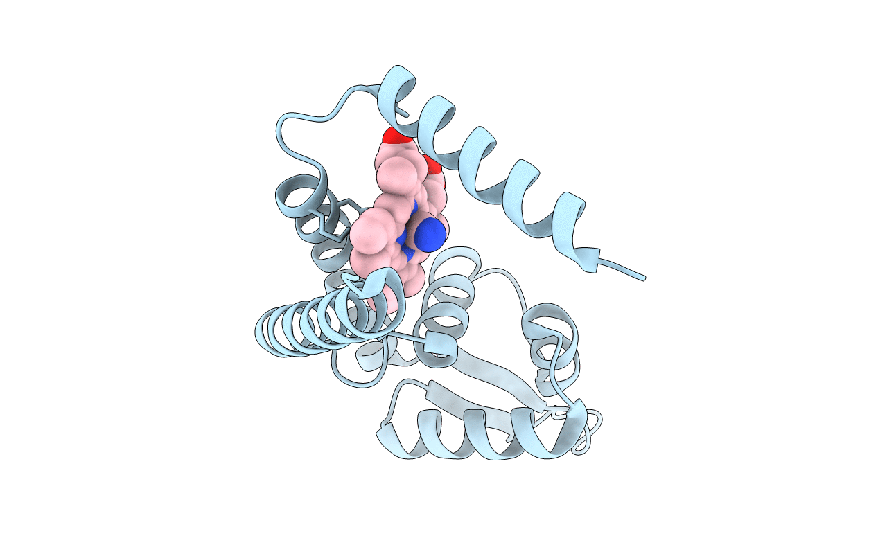
Title:
Crystal structure of heme sensor protein PefR in complex with heme and cyanide
Biological Source:
Source Organism(s):
Expression System(s):
Method Details:
Experimental Method:
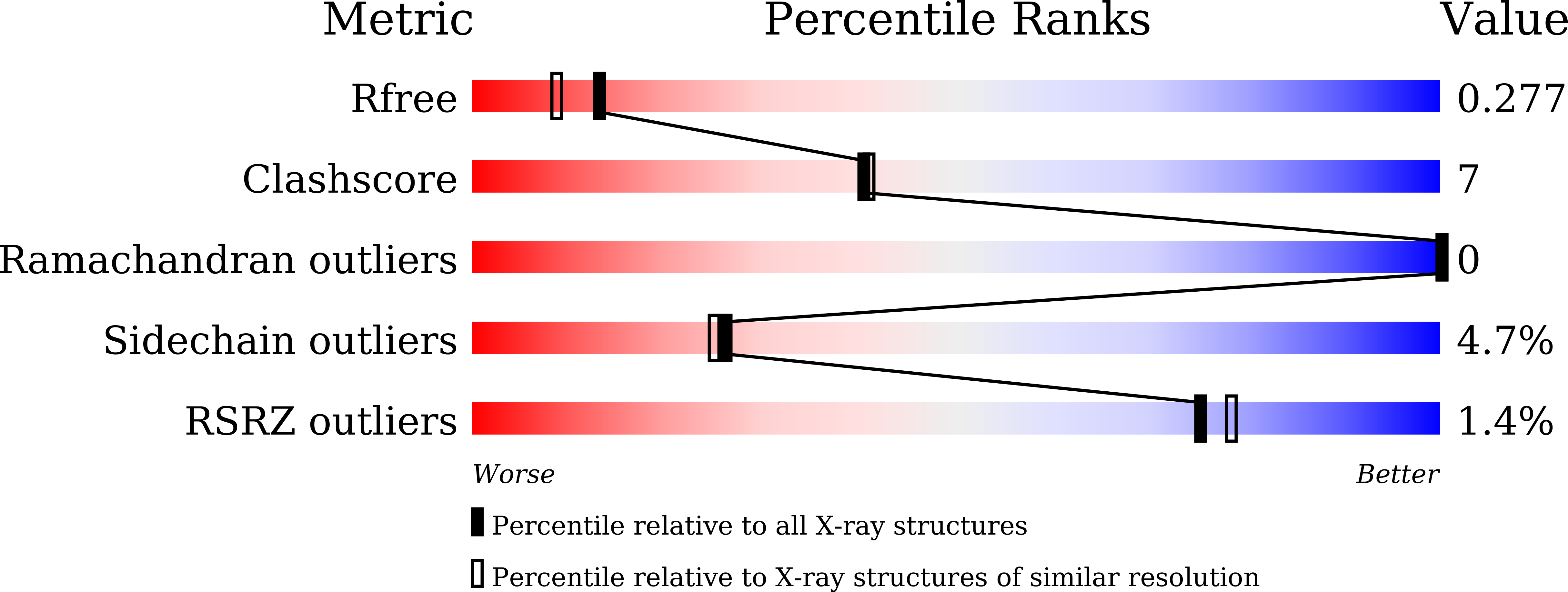
Resolution:
2.10 Å
R-Value Free:
0.27
R-Value Work:
0.22
R-Value Observed:
0.22
Space Group:
C 1 2 1