Deposition Date
2021-01-13
Release Date
2021-04-28
Last Version Date
2023-11-29
Entry Detail
PDB ID:
7DVL
Keywords:
Title:
Crystal Structure of the Catalytic Domain of Botulinum Neurotoxin Subtype A3
Biological Source:
Source Organism(s):
Clostridium botulinum (Taxon ID: 1491)
Expression System(s):
Method Details:
Experimental Method:
Resolution:
2.01 Å
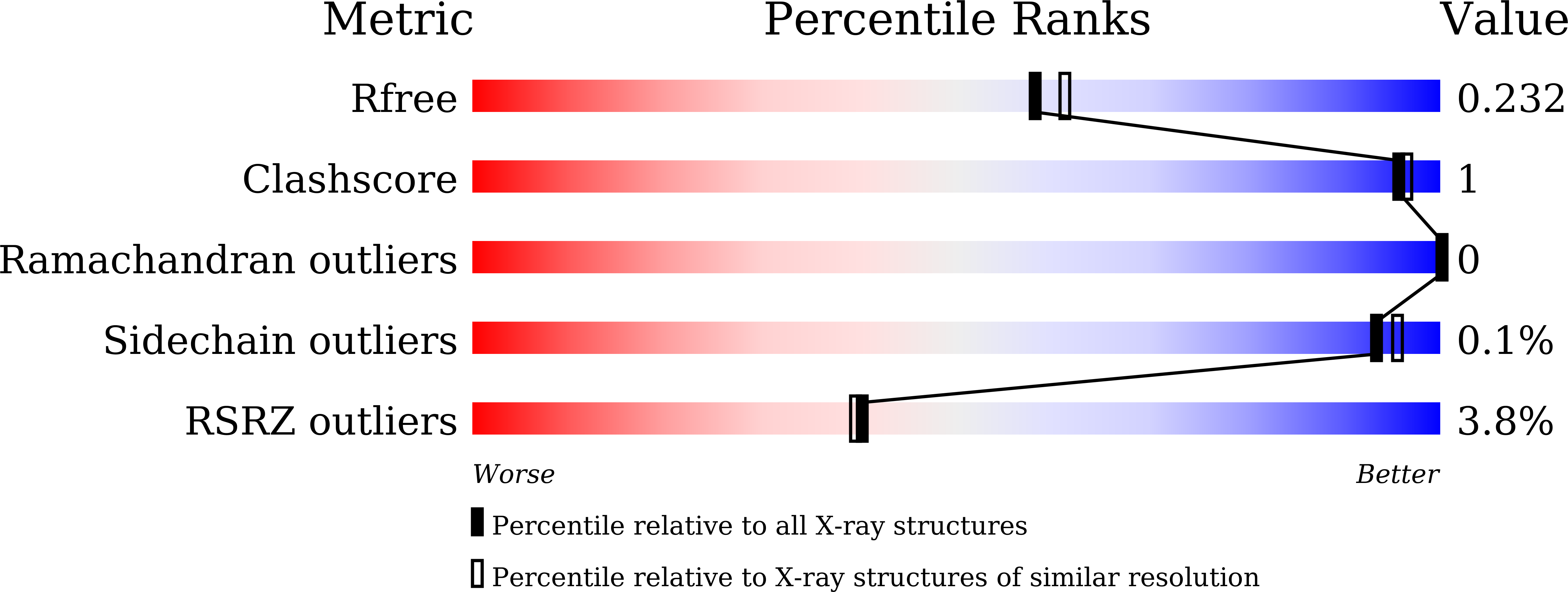
R-Value Free:
0.23
R-Value Work:
0.20
R-Value Observed:
0.20
Space Group:
P 21 21 21