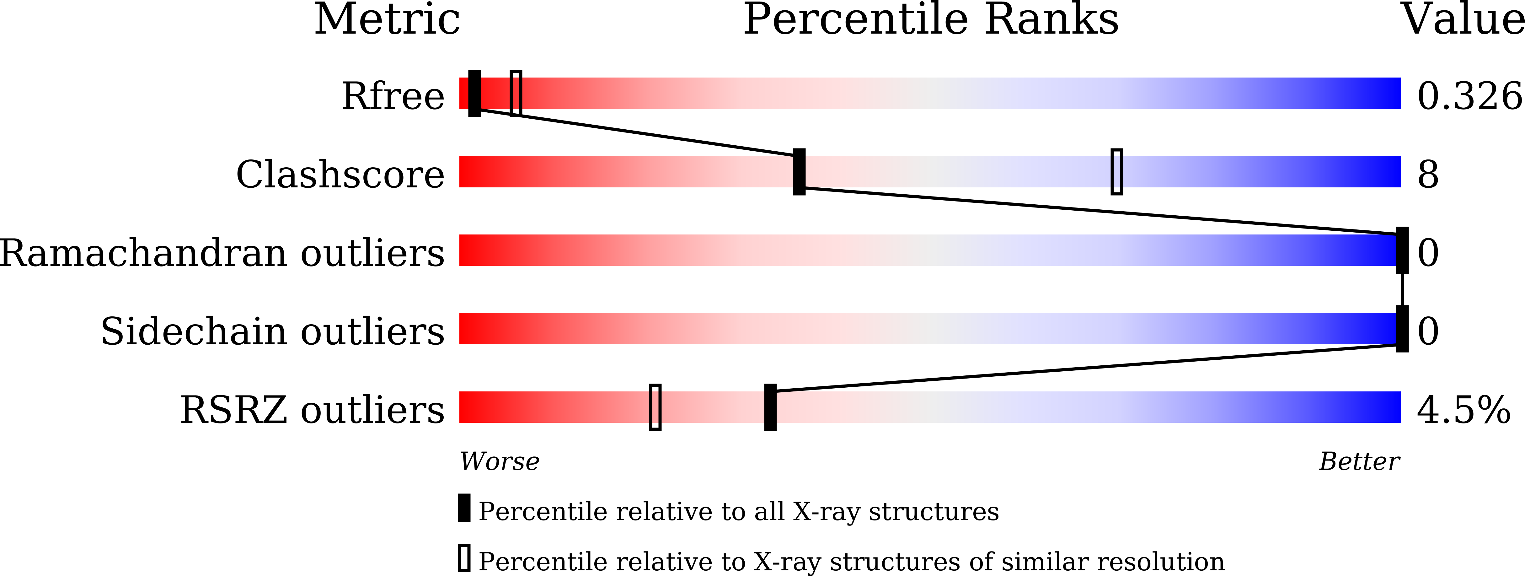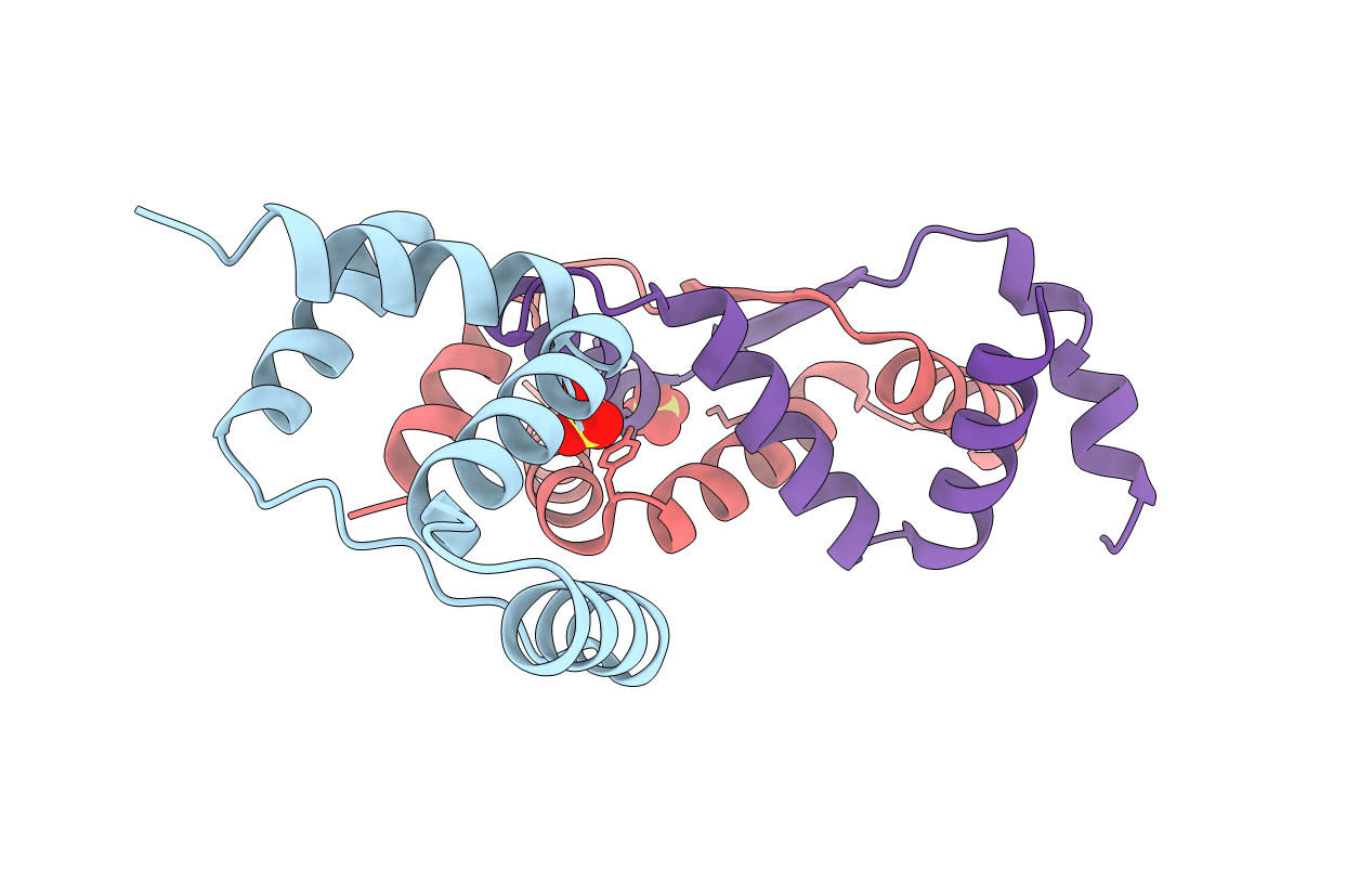
7DUV image
Deposition Date 2021-01-11
Release Date 2021-12-22
Last Version Date 2024-05-29
Entry Detail
PDB ID:
7DUV
Title:
Structure of Sulfolobus solfataricus SegB protein
Biological Source:
Method Details:
Experimental Method:
Resolution:
3.20 Å
R-Value Free:
0.32
R-Value Work:
0.28
R-Value Observed:
0.28
Space Group:
P 43 21 2
Macromolecular Entities
Polymer Type:polypeptide(L)
Molecule:SegB
Gene (Uniprot):SSO0035
Chain IDs:A, B, C
Chain Length:115
Number of Molecules:3
Biological Source:Saccharolobus solfataricus (strain ATCC 35092 / DSM 1617 / JCM 11322 / P2)
Ligand Molecules
Primary Citation
Chromosome segregation in Archaea: SegA- and SegB-DNA complex structures provide insights into segrosome assembly.
Nucleic Acids Res. 49 13150 13164 (2021)
PMID: 34850144 DOI: 10.1093/nar/gkab1155

Abstact

Genome segregation is a vital process in all organisms. Chromosome partitioning remains obscure in Archaea, the third domain of life. Here, we investigated the SegAB system from Sulfolobus solfataricus. SegA is a ParA Walker-type ATPase and SegB is a site-specific DNA-binding protein. We determined the structures of both proteins and those of SegA-DNA and SegB-DNA complexes. The SegA structure revealed an atypical, novel non-sandwich dimer that binds DNA either in the presence or in the absence of ATP. The SegB structure disclosed a ribbon-helix-helix motif through which the protein binds DNA site specifically. The association of multiple interacting SegB dimers with the DNA results in a higher order chromatin-like structure. The unstructured SegB N-terminus plays an essential catalytic role in stimulating SegA ATPase activity and an architectural regulatory role in segrosome (SegA-SegB-DNA) formation. Electron microscopy results also provide a compact ring-like segrosome structure related to chromosome organization. These findings contribute a novel mechanistic perspective on archaeal chromosome segregation.

Legend

Protein

Chemical

Disease

Primary Citation of related structures