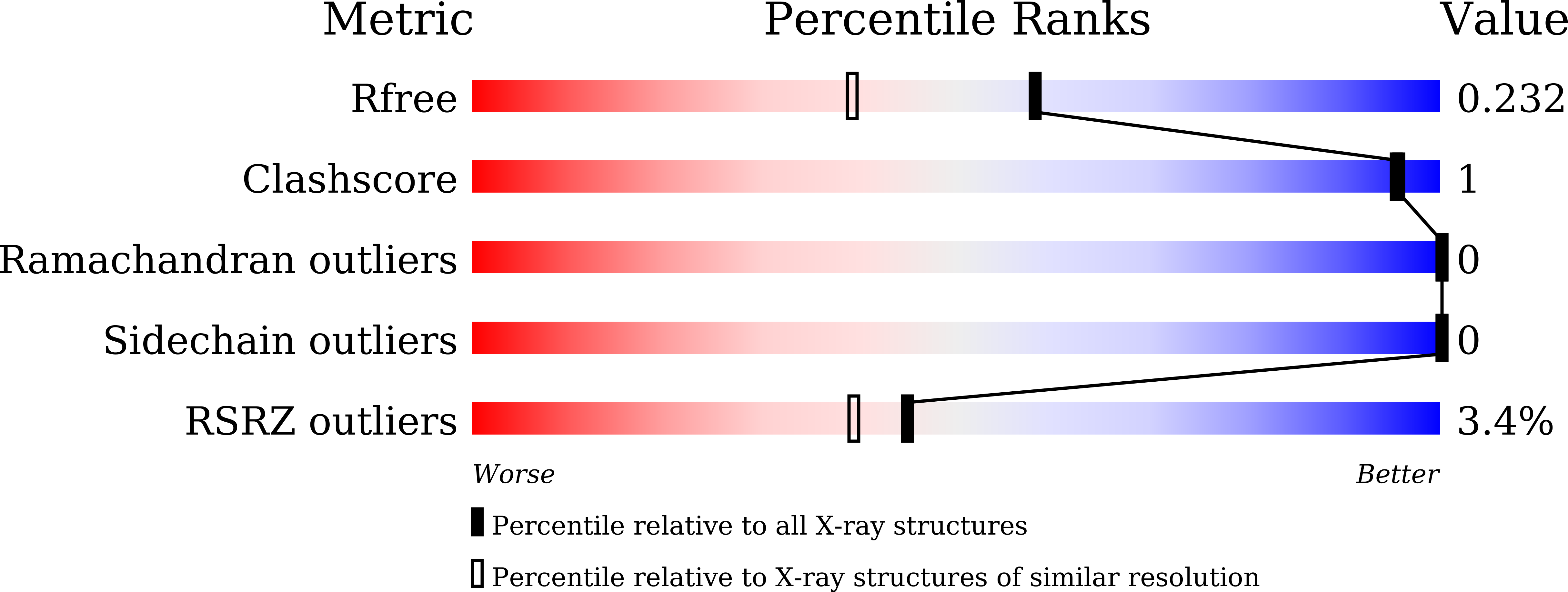
7DPY image
Entry Detail
PDB ID:
7DPY
Title:
Structure of Brucella abortus PhiA
Biological Source:
PDB Version:
Deposition Date:
2020-12-22
Release Date:
2021-06-23
Method Details:
Experimental Method:
Resolution:
1.80 Å
R-Value Free:
0.23
R-Value Work:
0.19
R-Value Observed:
0.20
Space Group:
P 21 21 2
Macromolecular Entities
Polymer Type:polypeptide(L)
Description:Brucella Abortus PhiA
Chain IDs:A, B
Chain Length:109
Number of Molecules:2
Biological Source:Brucella abortus bv. 1 str. 9-941
Primary Citation
Structure and Function of the Autolysin SagA in the Type IV Secretion System of Brucella abortus .
Mol.Cells 44 517 528 (2021)
PMID: 34112742 DOI: 10.14348/molcells.2021.0011

Abstact

A recent genetic study with Brucella abortus revealed the secretion activator gene A (SagA) as an autolysin component creating pores in the peptidoglycan (PGN) layer for the type IV secretion system (T4SS) and peptidoglycan hydrolase inhibitor A (PhiA) as an inhibitor of SagA. In this study, we determined the crystal structures of both SagA and PhiA. Notably, the SagA structure contained a PGN fragment in a space between the N- and C-terminal domains, showing the substrate-dependent hinge motion of the domains. The purified SagA fully hydrolyzed the meso-diaminopimelic acid (DAP)-type PGN, showing a higher activity than hen egg-white lysozyme. The PhiA protein exhibiting tetrameric assembly failed to inhibit SagA activity in our experiments. Our findings provide implications for the molecular basis of the SagA-PhiA system of B. abortus. The development of inhibitors of SagA would further contribute to controlling brucellosis by attenuating the function of T4SS, the major virulence factor of Brucella.

Legend

Protein

Chemical

Disease

Primary Citation of related structures