7DO7 image
Deposition Date 2020-12-12
Release Date 2021-02-03
Last Version Date 2024-03-27
Entry Detail
PDB ID:
7DO7
Keywords:
Title:
Crystal structure of Azotobacter vinelandii L-rhamnose 1-dehydrogenase(NAD and L-rhamnose bound-form)
Biological Source:
Method Details:
Experimental Method:
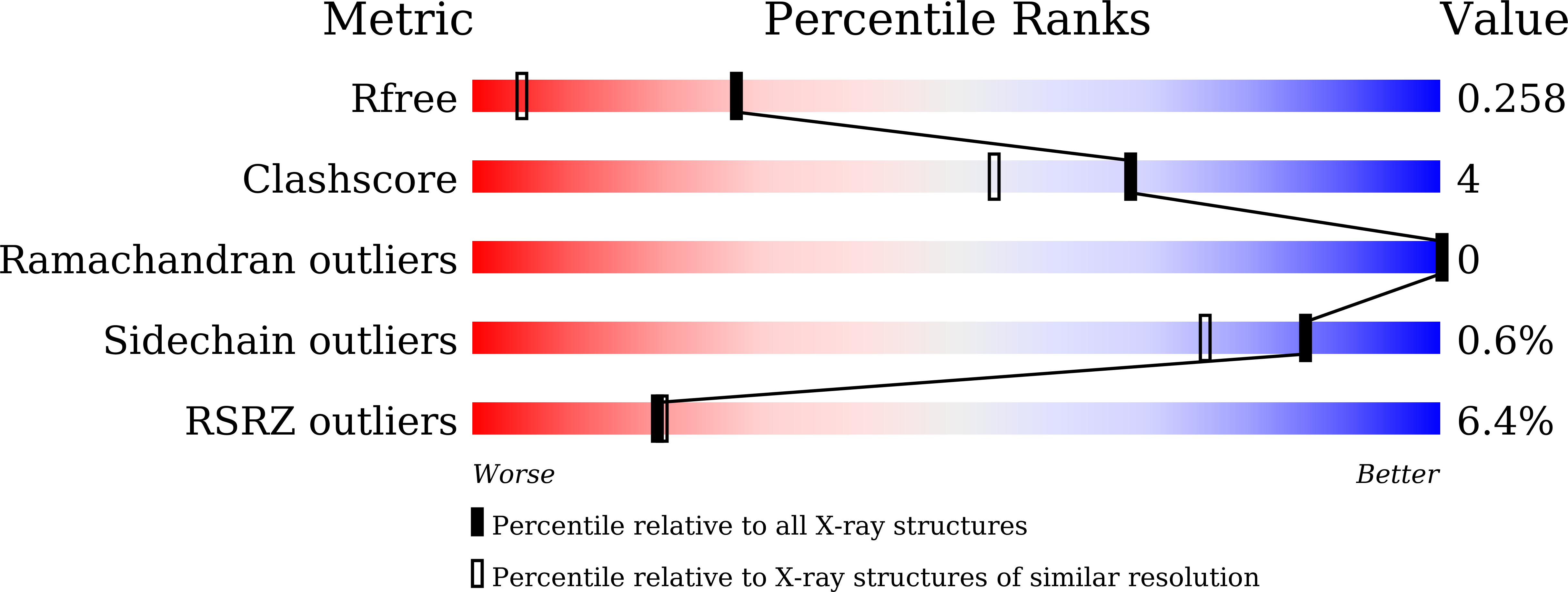
Resolution:
1.57 Å
R-Value Free:
0.25
R-Value Work:
0.22
R-Value Observed:
0.22
Space Group:
P 21 21 2
Macromolecular Entities
Polymer Type:polypeptide(L)
Molecule:Short-chain dehydrogenase/reductase SDR
Gene (Uniprot):LRA1
Chain IDs:A, B
Chain Length:267
Number of Molecules:2
Biological Source:Azotobacter vinelandii (strain DJ / ATCC BAA-1303)
Primary Citation
Crystal structure of l-rhamnose 1-dehydrogenase involved in the nonphosphorylative pathway of l-rhamnose metabolism in bacteria.
Febs Lett. 595 637 646 (2021)
PMID: 33482017 DOI: 10.1002/1873-3468.14046

Abstact

Several microorganisms can utilize l-rhamnose as a carbon and energy source through the nonphosphorylative metabolic pathway, in which l-rhamnose 1-dehydrogenase (RhaDH) catalyzes the NAD(P)+ -dependent oxidization of l-rhamnose to l-rhamnono-1,4-lactone. We herein investigated the crystal structures of RhaDH from Azotobacter vinelandii in ligand-free, NAD+ -bound, NADP+ -bound, and l-rhamnose- and NAD+ -bound forms at 1.9, 2.1, 2.4, and 1.6 Å resolution, respectively. The significant interactions with the 2'-phosphate group of NADP+ , but not the 2'-hydroxyl group of NAD+ , were consistent with a preference for NADP+ over NAD+ . The C5-OH and C6-methyl groups of l-rhamnose were recognized by specific residues of RhaDH through hydrogen bonds and hydrophobic contact, respectively, which contribute to the different substrate specificities from other aldose 1-dehydrogenases in the short-chain dehydrogenase/reductase superfamily.

Legend

Protein

Chemical

Disease

Primary Citation of related structures