7DN2 image
Entry Detail
PDB ID:
7DN2
EMDB ID:
Keywords:
Title:
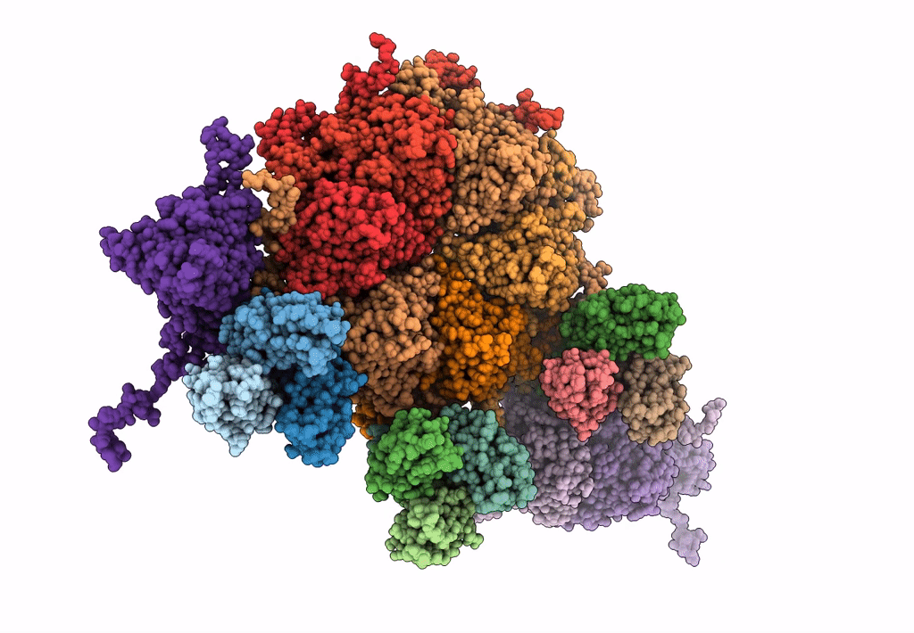
Acidic stable capsid structure of Helicobacter pylori bacteriophage KHP30
Biological Source:
PDB Version:
Deposition Date:
2020-12-08
Release Date:
2021-10-27
Method Details:
Experimental Method:
Resolution:
2.70 Å
Aggregation State:
PARTICLE
Reconstruction Method:
SINGLE PARTICLE
Macromolecular Entities
Polymer Type:polypeptide(L)
Description:Cement protein gp15
Chain IDs:J (auth: 1), K (auth: 2), L (auth: 3), M (auth: 4), N (auth: 5), O (auth: 6), P (auth: 7), Q (auth: 8), R (auth: 9)
Chain Length:126
Number of Molecules:9
Biological Source:Helicobacter pylori bacteriophage KHP30
Polymer Type:polypeptide(L)
Description:Major structural protein ORF14
Chain IDs:A (auth: a), B (auth: b), C (auth: c), D (auth: d), E (auth: e), F (auth: f), G (auth: g), H (auth: h), I (auth: i)
Chain Length:381
Number of Molecules:9
Biological Source:Helicobacter pylori bacteriophage KHP30
Ligand Molecules
Primary Citation
Acid-stable capsid structure of Helicobacter pylori bacteriophage KHP30 by single-particle cryoelectron microscopy.
Structure 30 300 ? (2022)
PMID: 34597601 DOI: 10.1016/j.str.2021.09.001

Abstact

The acid-stable capsid structures of Helicobacter pylori phages KHP30 and KHP40 are solved at 2.7 and 3.0 Å resolutions by cryoelectron microscopy, respectively. The capsids have icosahedral T = 9 symmetry and consist of each 540 copies of 2 structural proteins, a major capsid protein, and a cement protein. The major capsid proteins form 12 pentagonal capsomeres occupying icosahedral vertexes and 80 hexagonal capsomeres located at icosahedral faces and edges. The major capsid protein has a unique protruding loop extending to the neighboring subunit that stabilizes hexagonal capsomeres. Furthermore, the capsid is decorated with trimeric cement proteins with a jelly roll motif. The cement protein trimer sits on the quasi-three-fold axis formed by three major capsid protein capsomeres, thereby enhancing the particle stability by connecting these capsomeres. Sequence and structure comparisons between the related Helicobacter pylori phages suggest a possible mechanism of phage adaptation to the human gastric environment.

Legend

Protein

Chemical

Disease

Primary Citation of related structures