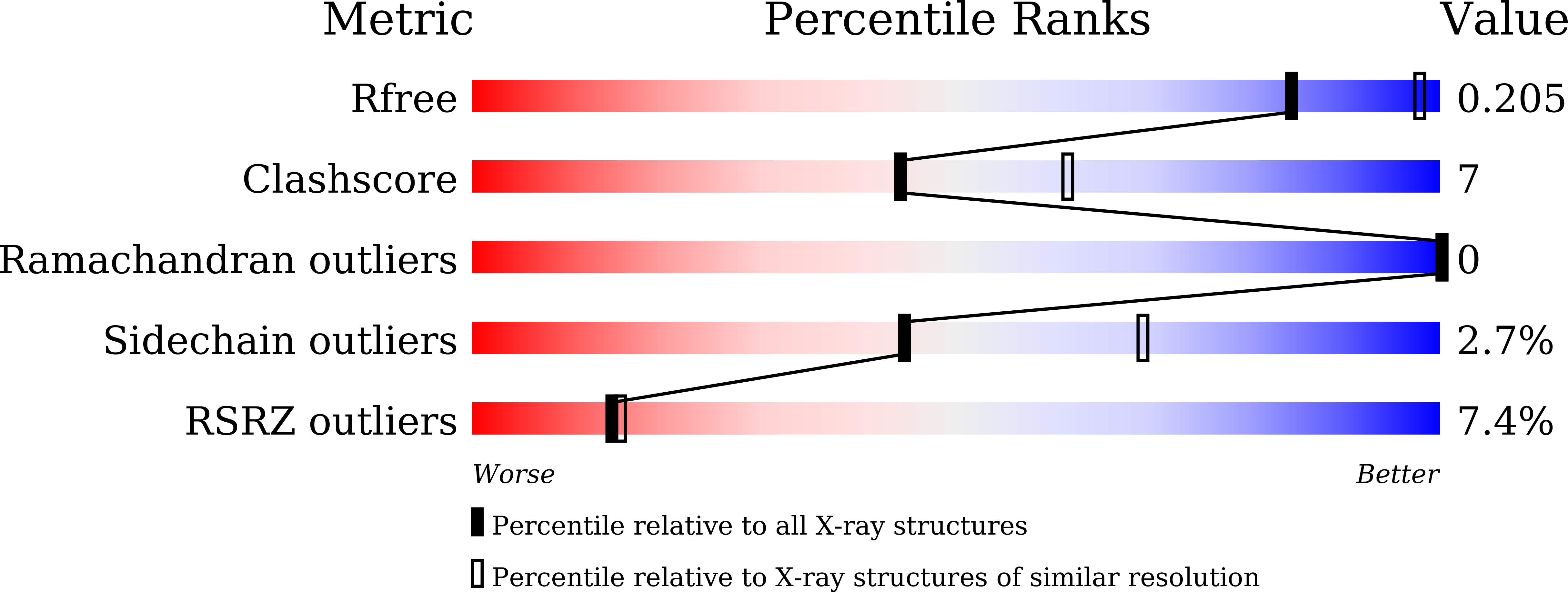
7DLV image
Deposition Date 2020-11-30
Release Date 2021-12-01
Last Version Date 2023-11-29
Entry Detail
PDB ID:
7DLV
Keywords:
Title:
shrimp dUTPase in complex with Stl
Biological Source:
Source Organism:
Host Organism:
Method Details:
Experimental Method:
Resolution:
2.53 Å
R-Value Free:
0.20
R-Value Work:
0.17
R-Value Observed:
0.17
Space Group:
P 1 21 1
Macromolecular Entities
Structures with similar UniProt ID
Protein Blast
Polymer Type:polypeptide(L)
Molecule:shrimp dUTPase
Chain IDs:A, B, C, G, H, I
Chain Length:149
Number of Molecules:6
Biological Source:Penaeus vannamei
Structures with similar UniProt ID
Protein Blast
Polymer Type:polypeptide(L)
Molecule:Orf20
Chain IDs:D, E, F, J, K, L
Chain Length:157
Number of Molecules:6
Biological Source:Staphylococcus aureus
Primary Citation
Structural basis of staphylococcal Stl inhibition on a eukaryotic dUTPase.
Int.J.Biol.Macromol. 184 821 830 (2021)
PMID: 34171258 DOI: 10.1016/j.ijbiomac.2021.06.107

Abstact

dUTPases are key enzymes in all life kingdoms. A staphylococcal repressor protein (Stl) inhibited dUTPases from multiple species to various extents. Understanding the molecular basis underlying the inhibition differences is crucial to develop effective proteinaceous inhibitors of dUTPases. Herein, we report the complex structure of Stl N-terminal domain (StlN-ter) and Litopenaeus vannamei dUTPase domain (lvDUT65-210). Stl inhibited lvDUT65-210 through its N-terminal domain. The lvDUT65-210-StlN-ter complex structure revealed a heterohexamer encompassing three StlN-ter monomers bound to one lvDUT65-210 trimer, generating two types of Stl-dUTPase interfaces. Interface I is formed by Stl interaction with the lvDUT65-210 active-site region that is contributed by motifs I-IV from its two subunits; interface II results from Stl binding to the C-terminal motif V of the third lvDUT65-210 subunit. Structural comparison revealed both conserved features and obvious differences in Stl-dUTPase interaction patterns, giving clues about the inhibition differences of Stl on dUTPases. Noticeably, interface II is only observed in lvDUT65-210-StlN-ter. The Stl-interacting residues of lvDUT65-210 are conserved in other eukaryotic dUTPases, particularly human dUTPase. Altogether, our study presents the first structural model of Stl interaction with eukaryotic dUTPase, contributing to a more complete view of Stl inhibition and facilitating the development of proteinaceous inhibitor for eukaryotic dUTPases.

Legend

Protein

Chemical

Disease

Primary Citation of related structures