7DG5 image
Deposition Date 2020-11-11
Release Date 2021-11-17
Last Version Date 2023-11-29
Entry Detail
PDB ID:
7DG5
Title:
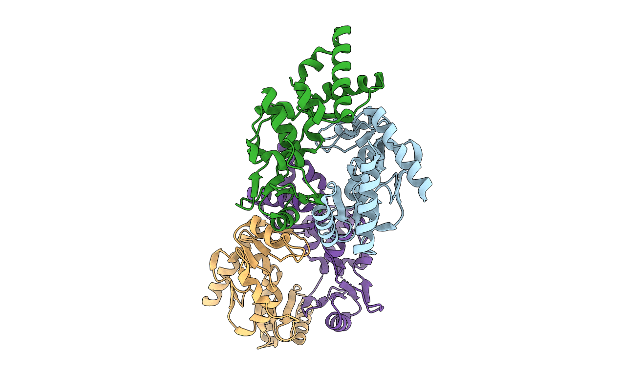
Crystal structure of mouse Smc1-Smc3 hinge domain containing a D574Y mutation
Biological Source:
Source Organism:
Mus musculus (Taxon ID: 10090)
Host Organism:
Method Details:
Experimental Method:
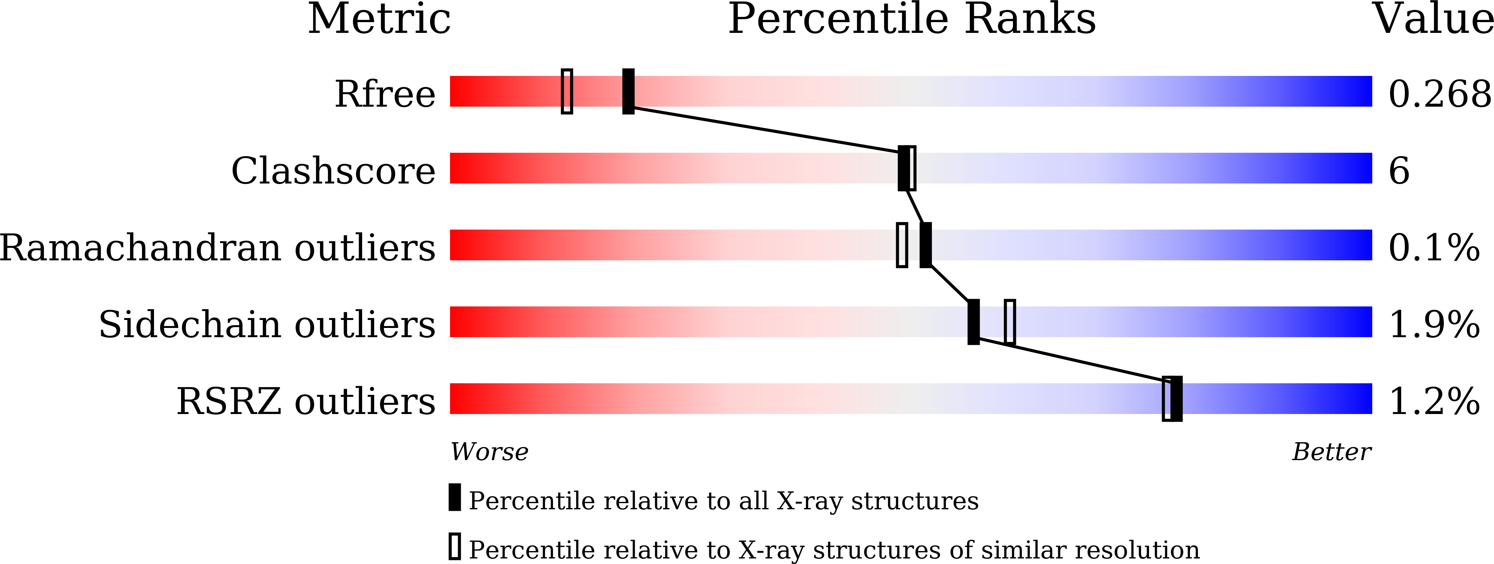
Resolution:
2.00 Å
R-Value Free:
0.26
R-Value Work:
0.21
R-Value Observed:
0.22
Space Group:
P 1
Macromolecular Entities
Polymer Type:polypeptide(L)
Molecule:Structural maintenance of chromosomes protein 1A
Gene (Uniprot):Smc1a
Mutations:D574Y
Chain IDs:A, C
Chain Length:215
Number of Molecules:2
Biological Source:Mus musculus
Polymer Type:polypeptide(L)
Molecule:Structural maintenance of chromosomes protein 3
Gene (Uniprot):Smc3
Chain IDs:B, D
Chain Length:213
Number of Molecules:2
Biological Source:Mus musculus
Primary Citation
Folding of cohesin's coiled coil is important for Scc2/4-induced association with chromosomes.
Elife 10 ? ? (2021)
PMID: 34259632 DOI: 10.7554/eLife.67268

Abstact

Cohesin's association with and translocation along chromosomal DNAs depend on an ATP hydrolysis cycle driving the association and subsequent release of DNA. This involves DNA being 'clamped' by Scc2 and ATP-dependent engagement of cohesin's Smc1 and Smc3 head domains. Scc2's replacement by Pds5 abrogates cohesin's ATPase and has an important role in halting DNA loop extrusion. The ATPase domains of all SMC proteins are separated from their hinge dimerisation domains by 50-nm-long coiled coils, which have been observed to zip up along their entire length and fold around an elbow, thereby greatly shortening the distance between hinges and ATPase heads. Whether folding exists in vivo or has any physiological importance is not known. We present here a cryo-EM structure of the apo form of cohesin that reveals the structure of folded and zipped-up coils in unprecedented detail and shows that Scc2 can associate with Smc1's ATPase head even when it is fully disengaged from that of Smc3. Using cysteine-specific crosslinking, we show that cohesin's coiled coils are frequently folded in vivo, including when cohesin holds sister chromatids together. Moreover, we describe a mutation (SMC1D588Y) within Smc1's hinge that alters how Scc2 and Pds5 interact with Smc1's hinge and that enables Scc2 to support loading in the absence of its normal partner Scc4. The mutant phenotype of loading without Scc4 is only explicable if loading depends on an association between Scc2/4 and cohesin's hinge, which in turn requires coiled coil folding.

Legend

Protein

Chemical

Disease

Primary Citation of related structures