Abstact
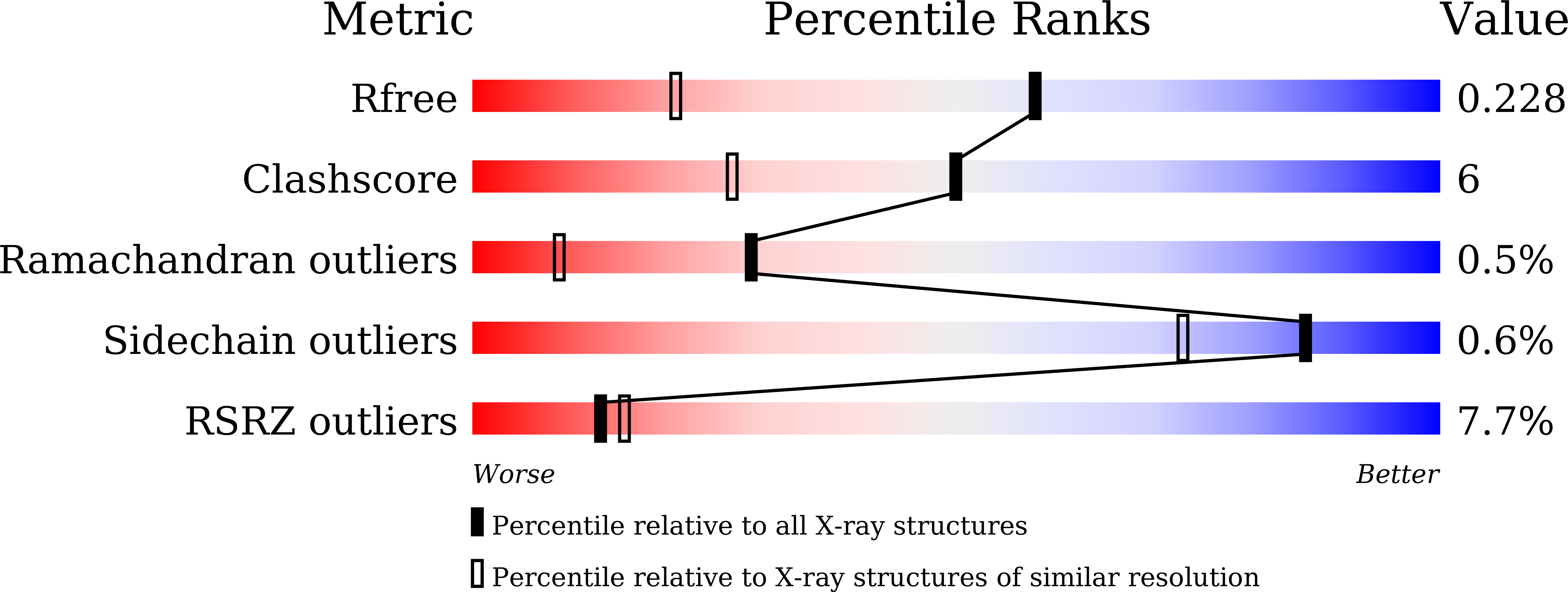
Lactoperoxidase (LPO) is a heme containing oxido-reductase enzyme. It is secreted from mammary, salivary, lachrymal and mucosal glands. It catalyses the conversion of thiocyanate into hypothiocyanate and halides into hypohalides. LPO belongs to the superfamily of mammalian heme peroxidases which also includes myeloperoxidase (MPO), eosinophil peroxidase (EPO) and thyroid peroxidase (TPO). The heme prosthetic group is covalently linked in LPO through two ester bonds involving conserved residues Glu258 and Asp108. It was isolated from colostrum of yak (Bos grunniens), purified to homogeneity and crystallized using ammonium iodide as a precipitating agent. The crystals belonged to monoclinic space group P21 with cell dimensions of a = 53.91 Å, b = 78.98 Å, c = 67.82 Å and β = 92.96°. The structure was determined at 1.55 Å resolution. This is the first structure of LPO from yak. Also, this is the highest resolution structure of LPO determined so far from any source. The structure determination revealed that three segments (Ser1-Cys15), (Thr117-Asn138) and (Cys167-Leu175) were disordered and formed one surface of LPO structure. In the substrate binding site, the iodide ions were observed in three subsites which are formed by (1) heme moiety and residues, Gln105, Asp108, His109, Phe113, Arg255, Glu258, Phe380 and Phe381, (2) residues, Asn230, Lys232, Pro236, Cys248, Phe254, Phe381 and Pro424 and (3) residues, Ser198, Leu199 and Arg202. The structure determination also revealed that the side chain of Phe254 was disordered. It was observed to adopt two conformations in the structures of LPO.


