
Deposition Date
2020-10-27
Release Date
2021-07-14
Last Version Date
2023-11-29
Entry Detail
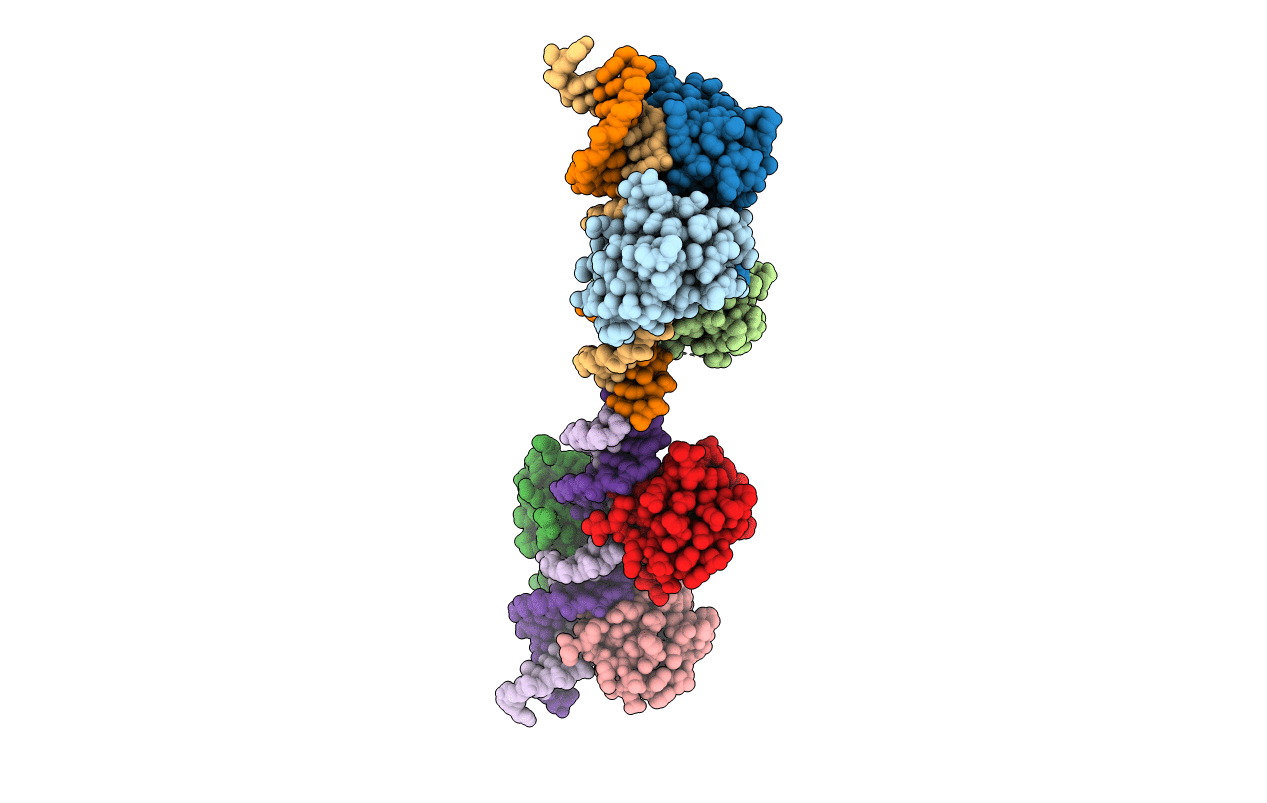
PDB ID:
7DCT
Keywords:
Title:
Crystal structure of HSF1 DNA-binding domain in complex with 3-site HSE DNA (24 bp)
Biological Source:
Source Organism(s):
Homo sapiens (Taxon ID: 9606)
Expression System(s):
Method Details:
Experimental Method:
Resolution:
2.36 Å
R-Value Free:
0.22
R-Value Work:
0.18
R-Value Observed:
0.18
Space Group:
P 21 21 21
