Deposition Date
2020-10-21
Release Date
2021-04-14
Last Version Date
2023-11-29
Entry Detail
PDB ID:
7DBS
Keywords:
Title:
Crystal Structure Of Biotin Protein Ligase From Leishmania Major in complex with Biotin
Biological Source:
Source Organism(s):
Leishmania major (Taxon ID: 5664)
Expression System(s):
Method Details:
Experimental Method:
Resolution:
2.33 Å
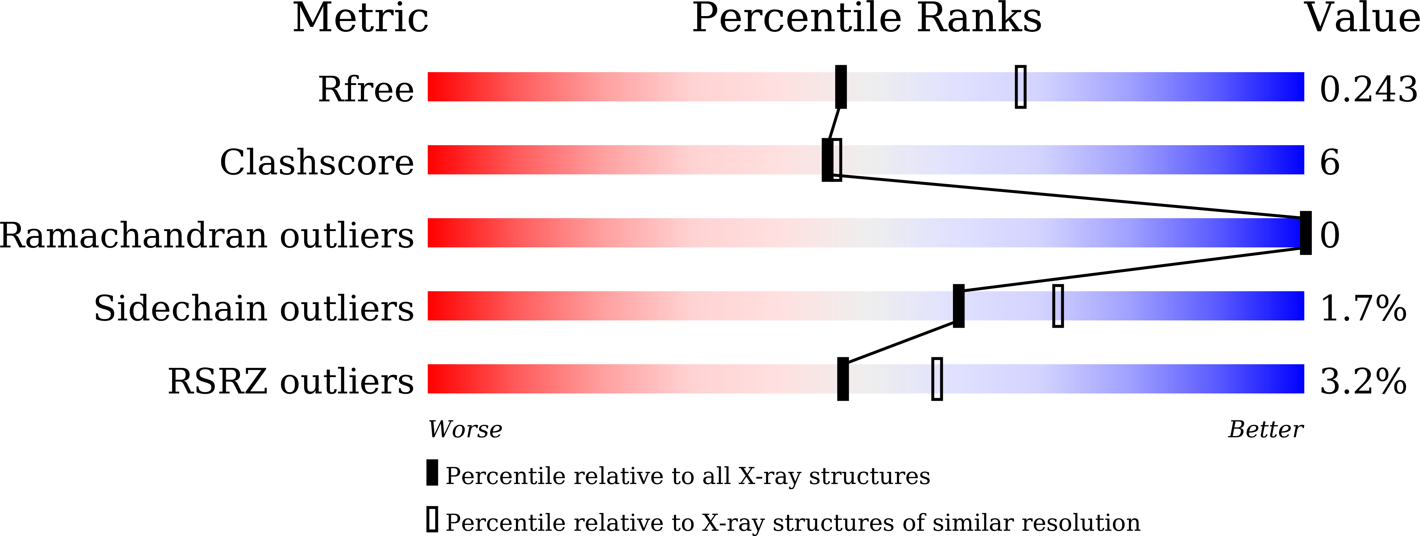
R-Value Free:
0.24
R-Value Work:
0.21
R-Value Observed:
0.21
Space Group:
C 1 2 1