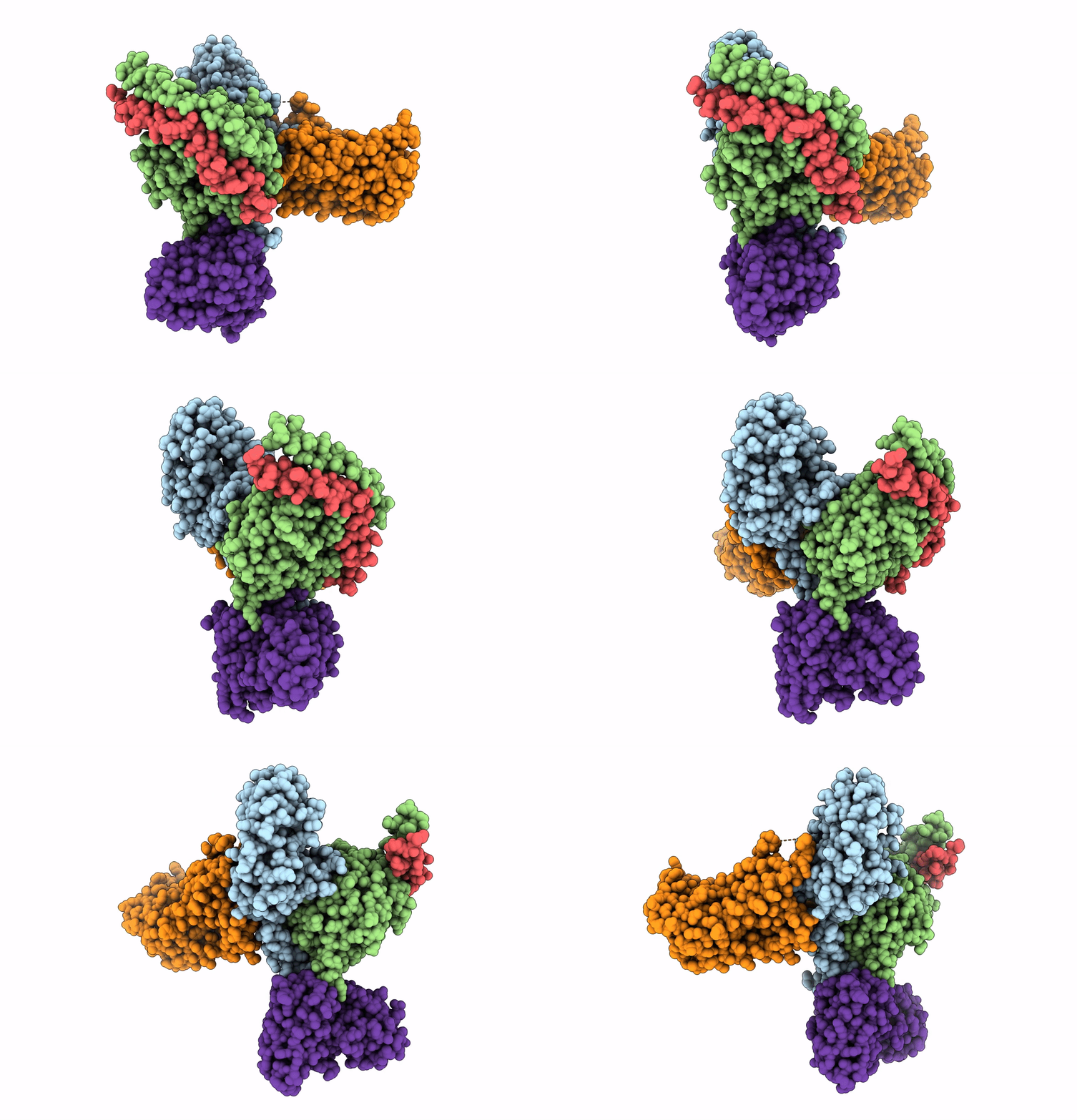
7DB6 image
Entry Detail
PDB ID:
7DB6
EMDB ID:
Title:
human melatonin receptor MT1 - Gi1 complex
Biological Source:
PDB Version:
Deposition Date:
2020-10-19
Release Date:
2021-08-18
Method Details:
Experimental Method:
Resolution:
3.30 Å
Aggregation State:
PARTICLE
Reconstruction Method:
SINGLE PARTICLE
Macromolecular Entities
Polymer Type:polypeptide(L)
Description:Guanine nucleotide-binding protein G(i) subunit alpha-1
Chain IDs:A
Chain Length:354
Number of Molecules:1
Biological Source:Homo sapiens
Polymer Type:polypeptide(L)
Description:Guanine nucleotide-binding protein G(I)/G(S)/G(T) subunit beta-1
Chain IDs:B
Chain Length:344
Number of Molecules:1
Biological Source:Rattus rattus
Polymer Type:polypeptide(L)
Description:Guanine nucleotide-binding protein G(I)/G(S)/G(O) subunit gamma-2
Chain IDs:C
Chain Length:68
Number of Molecules:1
Biological Source:Bos taurus
Polymer Type:polypeptide(L)
Description:Melatonin receptor type 1A
Chain IDs:E (auth: D)
Chain Length:372
Number of Molecules:1
Biological Source:Homo sapiens
Polymer Type:polypeptide(L)
Description:scFv16
Chain IDs:D (auth: E)
Chain Length:260
Number of Molecules:1
Biological Source:Mus musculus
Ligand Molecules
Primary Citation
Cryo-EM structure of the human MT 1 -G i signaling complex.
Nat.Struct.Mol.Biol. 28 694 701 (2021)
PMID: 34354246 DOI: 10.1038/s41594-021-00634-1

Abstact

Melatonin receptors (MT1 and MT2) transduce inhibitory signaling by melatonin (N-acetyl-5-methoxytryptamine), which is associated with sleep induction and circadian rhythm modulation. Although recently reported crystal structures of ligand-bound MT1 and MT2 elucidated the basis of ligand entry and recognition, the ligand-induced MT1 rearrangement leading to Gi-coupling remains unclear. Here we report a cryo-EM structure of the human MT1-Gi signaling complex at 3.3 Å resolution, revealing melatonin-induced conformational changes propagated to the G-protein-coupling interface during activation. In contrast to other Gi-coupled receptors, MT1 exhibits a large outward movement of TM6, which is considered a specific feature of Gs-coupled receptors. Structural comparison of Gi and Gs complexes demonstrated conformational diversity of the C-terminal entry of the Gi protein, suggesting loose and variable interactions at the end of the α5 helix of Gi protein. These notions, together with our biochemical and computational analyses, highlight variable binding modes of Gαi and provide the basis for the selectivity of G-protein signaling.

Legend

Protein

Chemical

Disease

Primary Citation of related structures