Deposition Date
2020-10-16
Release Date
2021-06-02
Last Version Date
2023-11-29
Entry Detail
PDB ID:
7DAN
Keywords:
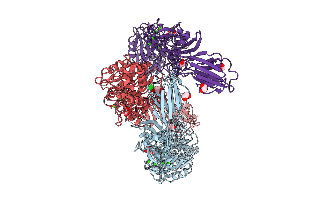
Title:
Structure of the Ca2+-bound wild-type peptidylarginine deiminase type III (PAD3)
Biological Source:
Source Organism(s):
Homo sapiens (Taxon ID: 9606)
Expression System(s):
Method Details:
Experimental Method:
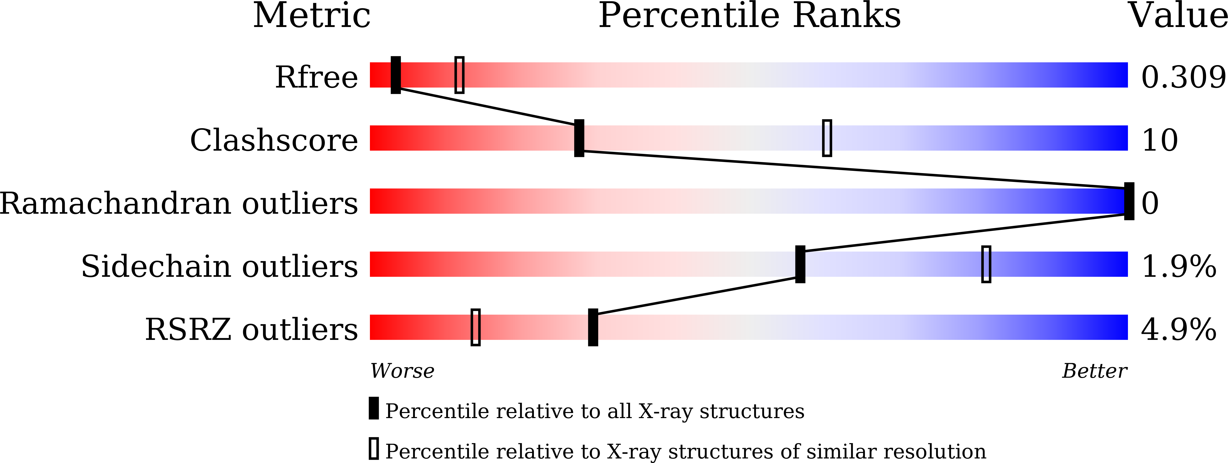
Resolution:
3.10 Å
R-Value Free:
0.30
R-Value Work:
0.23
R-Value Observed:
0.24
Space Group:
C 1 2 1