7DAD image
Deposition Date 2020-10-16
Release Date 2021-03-24
Last Version Date 2023-11-29
Entry Detail
PDB ID:
7DAD
Keywords:
Title:
EPD in complex with tubulin
Biological Source:
Source Organism:
Mus musculus (Taxon ID: 10090)
Gallus gallus (Taxon ID: 9031)
Sus scrofa (Taxon ID: 9823)
Host Organism:
Method Details:
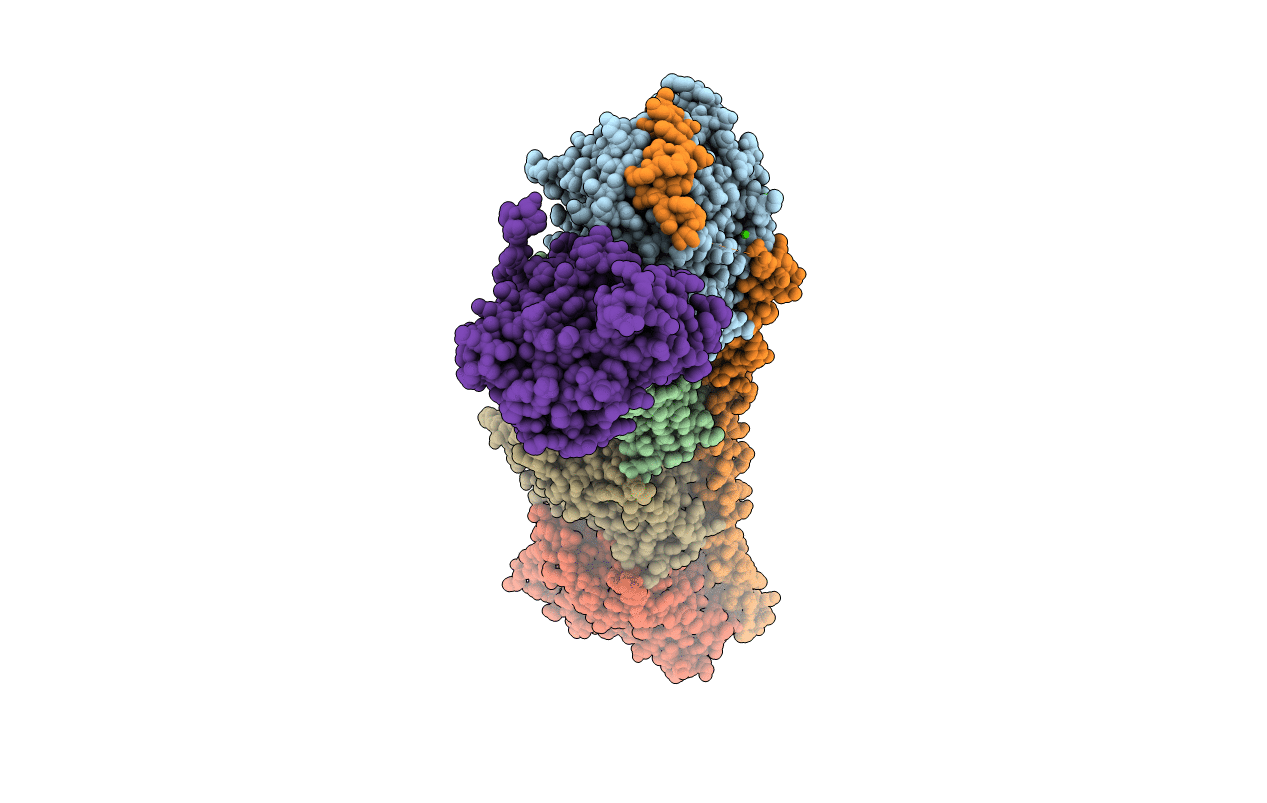
Experimental Method:
Resolution:
2.85 Å
R-Value Free:
0.22
R-Value Work:
0.17
R-Value Observed:
0.17
Space Group:
P 21 21 21
Macromolecular Entities
Structures with similar UniProt ID
Protein Blast
Polymer Type:polypeptide(L)
Molecule:Tubulin alpha-1B chain
Gene (Uniprot):TUBA1B
Chain IDs:A, C
Chain Length:451
Number of Molecules:2
Biological Source:Sus scrofa
Structures with similar UniProt ID
Protein Blast
Polymer Type:polypeptide(L)
Molecule:Tubulin beta chain
Chain IDs:B, D
Chain Length:445
Number of Molecules:2
Biological Source:Sus scrofa
Structures with similar UniProt ID
Protein Blast
Polymer Type:polypeptide(L)
Molecule:Stathmin-4
Gene (Uniprot):Stmn4
Chain IDs:E
Chain Length:143
Number of Molecules:1
Biological Source:Mus musculus
Polymer Type:polypeptide(L)
Molecule:Tubulin tyrosine ligase
Chain IDs:F
Chain Length:384
Number of Molecules:1
Biological Source:Gallus gallus
Primary Citation
High-resolution X-ray structure of three microtubule-stabilizing agents in complex with tubulin provide a rationale for drug design.
Biochem.Biophys.Res.Commun. 534 330 336 (2021)
PMID: 33272565 DOI: 10.1016/j.bbrc.2020.11.082

Abstact

Microtubule is a key component of cytoskeleton and has been considered as an important target for the treatment of cancer. In particular, the tubulin taxane-site inhibitors such as taxol analogs and epothilones have achieved great success in clinical trials. However, the structural basis of many taxane-site inhibitors is still lacking in exploring their mechanism of action. We here reported crystal complex structures for three taxane-site inhibitors, Ixabepilone, Epothilone B, and Epothilone D, which were determined to 2.4 Å, 2.4 Å, and 2.85 Å, respectively. The crystal structures revealed that these taxane-site inhibitors possess similar binding modes to that of Epothilone A at the taxane site, e.g. making critical hydrogen-bonding interactions with multiple residues on the M-loop, which facilitating the tubulin polymerization. Furthermore, we summarized the binding modes of almost all taxane-site inhibitors and identified novel taxane-site ligands with simpler chemical structures through virtual screening. On this basis, new derivatives with higher binding affinity to tubulin were designed and developed, which can form additional hydrogen bond interactions with tubulin. Overall, this work determined the mechanism of action of epothilones and provided a structural basis to design reasonably novel taxane-site inhibitors with simpler structure and improved pharmacokinetic properties.

Legend

Protein

Chemical

Disease

Primary Citation of related structures