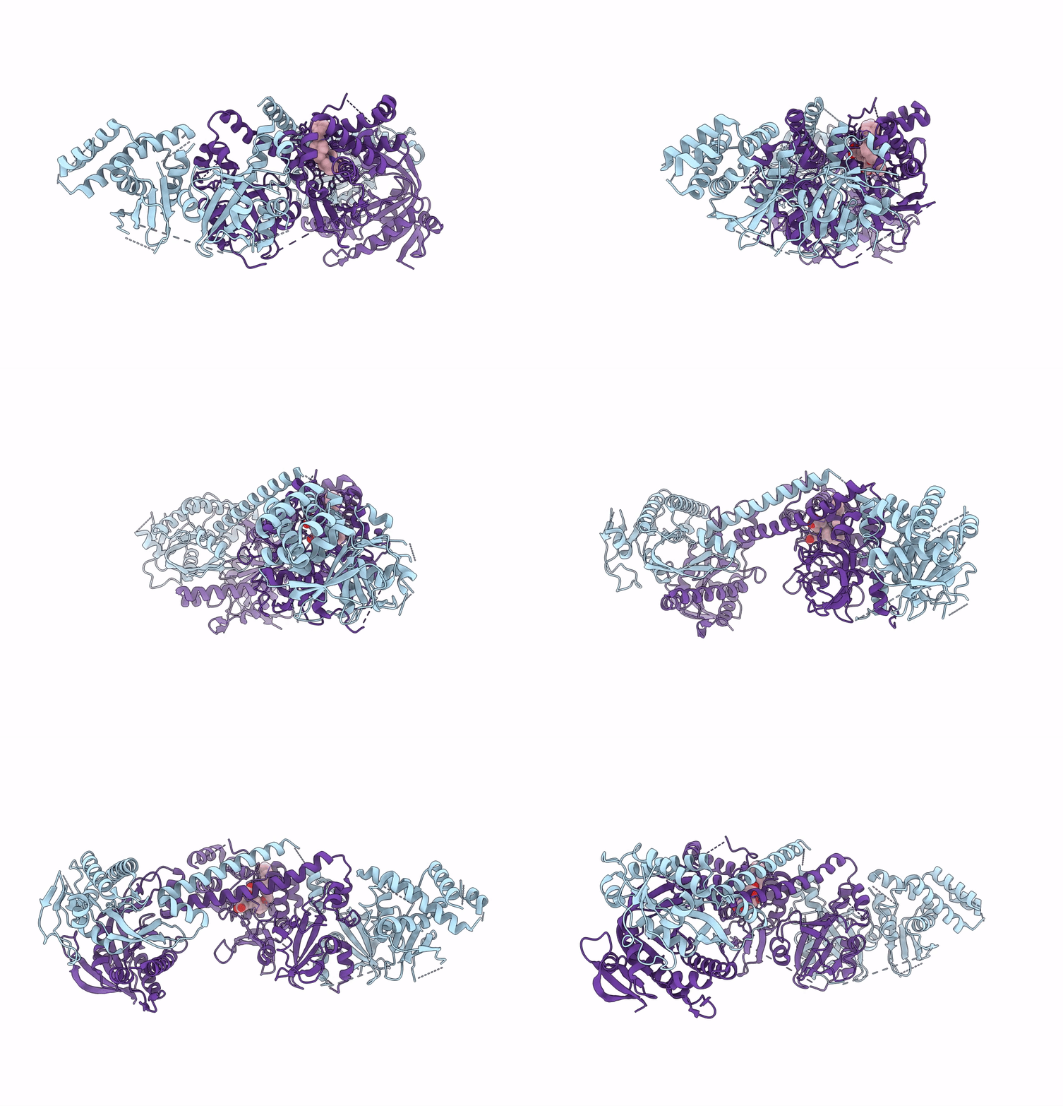
7D9T image
Deposition Date 2020-10-14
Release Date 2021-08-11
Last Version Date 2024-05-29
Entry Detail
PDB ID:
7D9T
Title:
Structure of human soluble guanylate cyclase in the cinciguat-bound inactive state
Biological Source:
Source Organism:
Homo sapiens (Taxon ID: 9606)
Host Organism:
Method Details:
Experimental Method:
Resolution:
4.10 Å
Aggregation State:
PARTICLE
Reconstruction Method:
SINGLE PARTICLE
Macromolecular Entities
Polymer Type:polypeptide(L)
Molecule:Guanylate cyclase soluble subunit alpha-1
Gene (Uniprot):GUCY1A1
Chain IDs:A
Chain Length:690
Number of Molecules:1
Biological Source:Homo sapiens
Polymer Type:polypeptide(L)
Molecule:Guanylate cyclase soluble subunit beta-1
Gene (Uniprot):GUCY1B1
Chain IDs:B
Chain Length:619
Number of Molecules:1
Biological Source:Homo sapiens
Ligand Molecules
Primary Citation
Activation mechanism of human soluble guanylate cyclase by stimulators and activators.
Nat Commun 12 5492 5492 (2021)
PMID: 34535643 DOI: 10.1038/s41467-021-25617-0

Abstact

Soluble guanylate cyclase (sGC) is the receptor for nitric oxide (NO) in human. It is an important validated drug target for cardiovascular diseases. sGC can be pharmacologically activated by stimulators and activators. However, the detailed structural mechanisms, through which sGC is recognized and positively modulated by these drugs at high spacial resolution, are poorly understood. Here, we present cryo-electron microscopy structures of human sGC in complex with NO and sGC stimulators, YC-1 and riociguat, and also in complex with the activator cinaciguat. These structures uncover the molecular details of how stimulators interact with residues from both β H-NOX and CC domains, to stabilize sGC in the extended active conformation. In contrast, cinaciguat occupies the haem pocket in the β H-NOX domain and sGC shows both inactive and active conformations. These structures suggest a converged mechanism of sGC activation by pharmacological compounds.

Legend

Protein

Chemical

Disease

Primary Citation of related structures