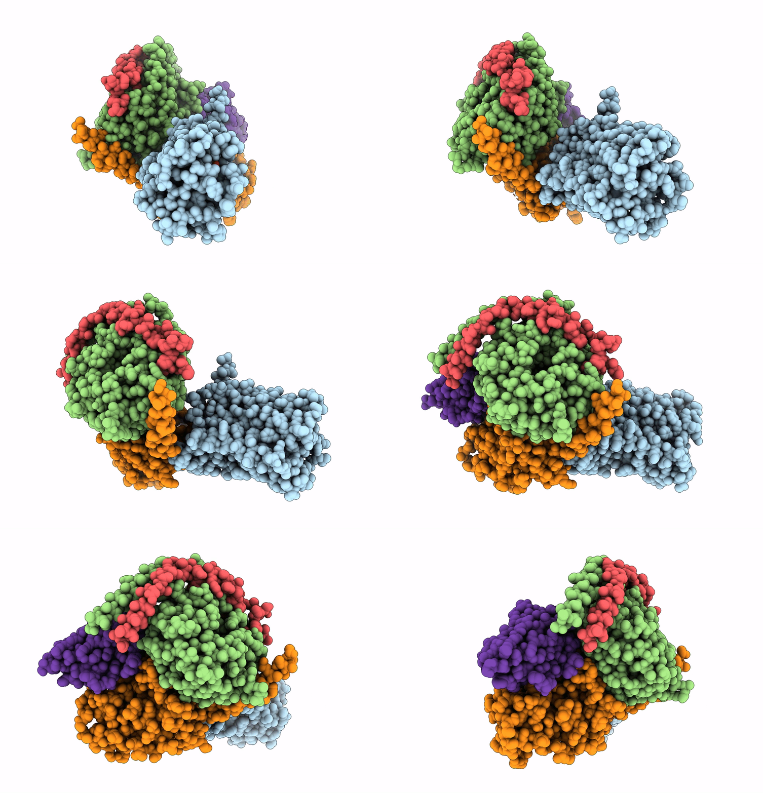
7D7M image
Entry Detail
PDB ID:
7D7M
EMDB ID:
Title:
Cryo-EM Structure of the Prostaglandin E Receptor EP4 Coupled to G Protein
Biological Source:
Source Organism:
Host Organism:
PDB Version:
Deposition Date:
2020-10-05
Release Date:
2020-11-18
Method Details:
Experimental Method:
Resolution:
3.30 Å
Aggregation State:
2D ARRAY
Reconstruction Method:
SINGLE PARTICLE
Macromolecular Entities
Polymer Type:polypeptide(L)
Description:Prostaglandin E2 receptor EP4 subtype,Prostaglandin E2 receptor EP4 subtype
Mutations:N7Q, N177Q, 218-259 deletion
Chain IDs:A
Chain Length:333
Number of Molecules:1
Biological Source:Homo sapiens
Polymer Type:polypeptide(L)
Description:Guanine nucleotide-binding protein G(I)/G(S)/G(T) subunit beta-1
Chain IDs:B
Chain Length:345
Number of Molecules:1
Biological Source:Homo sapiens
Polymer Type:polypeptide(L)
Description:Guanine nucleotide-binding protein G(I)/G(S)/G(O) subunit gamma-2
Chain IDs:C
Chain Length:71
Number of Molecules:1
Biological Source:Homo sapiens
Polymer Type:polypeptide(L)
Description:Guanine nucleotide-binding protein G(s) subunit alpha isoforms short,Guanine nucleotide-binding protein G(s) subunit alpha isoforms short,Guanine nucleotide-binding protein G(s) subunit alpha isoforms short
Mutations:65 to 203, 255 to 264 deletion G49D, E50N, L63Y, A249D, S252D, I372A and V375I
Chain IDs:D
Chain Length:249
Number of Molecules:1
Biological Source:Homo sapiens
Polymer Type:polypeptide(L)
Description:nanobody Nb35
Chain IDs:E
Chain Length:134
Number of Molecules:1
Biological Source:Lama glama
Ligand Molecules
Primary Citation
Cryo-EM Structure of the Prostaglandin E Receptor EP4 Coupled to G Protein.
Structure 29 252 260.e6 (2021)
PMID: 33264604 DOI: 10.1016/j.str.2020.11.007

Abstact

Prostaglandin E receptor EP4, a class A G protein-coupled receptor (GPCR), is a common drug target in various disorders, such as acute decompensated heart failure and ulcerative colitis. Here, we report the cryoelectron microscopy (cryo-EM) structure of the EP4-heterotrimeric G protein (Gs) complex with the endogenous ligand at a global resolution of 3.3 Å. In this structure, compared with that in the inactive EP4 structure, the sixth transmembrane domain is shifted outward on the intracellular side, although the shift is smaller than that in other class A GPCRs bound to Gs. Instead, the C-terminal helix of Gs is inserted toward TM2 of EP4, and the conserved C-terminal hook structure formsthe extended state. These structural features are formed by the conserved residues in prostanoid receptors (Phe542.39 and Trp3277.51). These findings may be important for the thorough understanding of the G protein-binding mechanism of EP4 and other prostanoid receptors.

Legend

Protein

Chemical

Disease

Primary Citation of related structures