7D6F image
Deposition Date 2020-09-30
Release Date 2020-11-04
Last Version Date 2023-11-29
Entry Detail
PDB ID:
7D6F
Title:
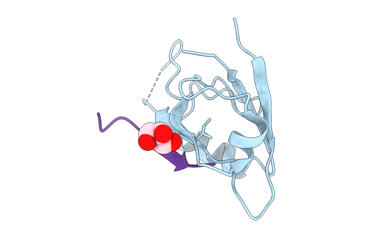
The crystal structure of ARMS-PBM/MAGI2-PDZ4
Biological Source:
Source Organism:
Mus musculus (Taxon ID: 10090)
Rattus norvegicus (Taxon ID: 10116)
Host Organism:
Method Details:
Experimental Method:
Resolution:
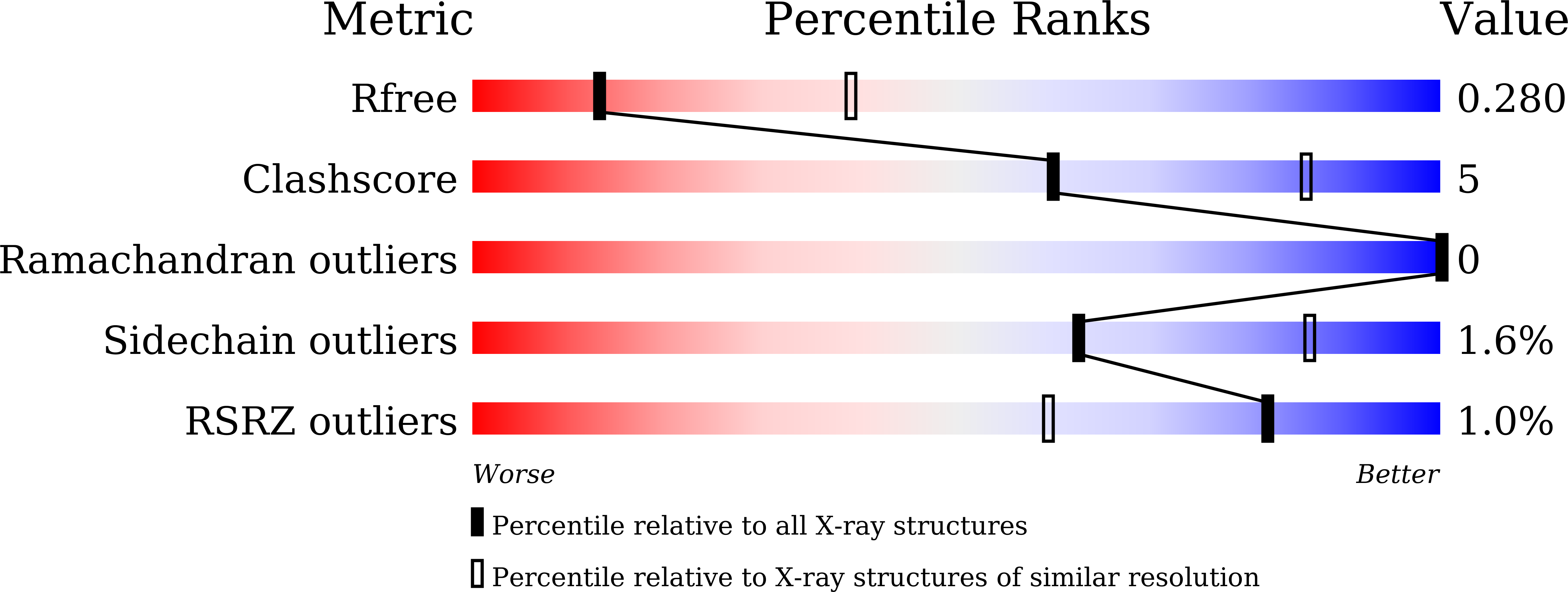
3.00 Å
R-Value Free:
0.28
R-Value Work:
0.23
R-Value Observed:
0.23
Space Group:
C 2 2 2
Macromolecular Entities
Structures with similar UniProt ID
Protein Blast
Polymer Type:polypeptide(L)
Molecule:Membrane-associated guanylate kinase, WW and PDZ domain-containing protein 2
Gene (Uniprot):Magi2
Chain IDs:A
Chain Length:101
Number of Molecules:1
Biological Source:Mus musculus
Structures with similar UniProt ID
Protein Blast
Polymer Type:polypeptide(L)
Molecule:Kinase D-interacting substrate of 220 kDa
Gene (Uniprot):Kidins220
Chain IDs:B
Chain Length:19
Number of Molecules:1
Biological Source:Rattus norvegicus
Ligand Molecules
Primary Citation
Crystal structure of the PDZ4 domain of MAGI2 in complex with PBM of ARMS reveals a canonical PDZ recognition mode.
Neurochem.Int. 149 105152 105152 (2021)
PMID: 34371146 DOI: 10.1016/j.neuint.2021.105152

Abstact

Membrane-associated guanylate kinase, WW and PDZ domain-containing protein 2 (MAGI2) is a neuronal scaffold protein that plays critical roles at synaptic junctions by assembling neurotransmitter receptors and cell adhesion proteins through its multiple protein-protein interaction domains, including six PDZ domains, two phosphoserine-phosphothreonine binding WW domains, and a guanylate kinase GK domain. Previous studies showed that MAGI2 participates in formation of tetrameric complexes with PDZ-GEF1, TrkA receptor, and ankyrin repeat-rich membrane spanning (ARMS) protein at late endosomes and is crucial for neurite outgrowth. However, the molecular mechanism governing the assembly of these complexes remains unknown. Here, we characterize the direct interaction between MAGI2 and ARMS through multiple biochemical assays. Moreover, our solved crystal structure of the truncated PDZ4/PBM (PDZ binding motifs) complex of MAGI2 and ARMS proteins (MAGI2-PDZ4/ARMS-PBM) reveals that the binding interface lies between the αB/βB groove from the PDZ4 of MAGI2 and the C-terminal PBM from ARMS. The structure reveals high similarity to others in this protein family where canonical PDZ/PBM interactions are observed. However, the conserved "GLGF" motif in the PSD-95-PDZ3 changes to "GFGF" in the MAGI2-PDZ4/ARMS-PBM complex. We further validated our crystal structure through serial mutagenesis assays. Taken together, our study provides the biochemical details and binding mechanisms that underpin the stabilization of the MAGI2-PDZ4/ARMS-PBM complex, thereby offering a biochemical and structural basis for further understanding of the functional roles of MAGI2, ARMS, PDZ-GEF1, and TrkA in forming the tetrameric receptor complex in neuronal signaling.

Legend

Protein

Chemical

Disease

Primary Citation of related structures