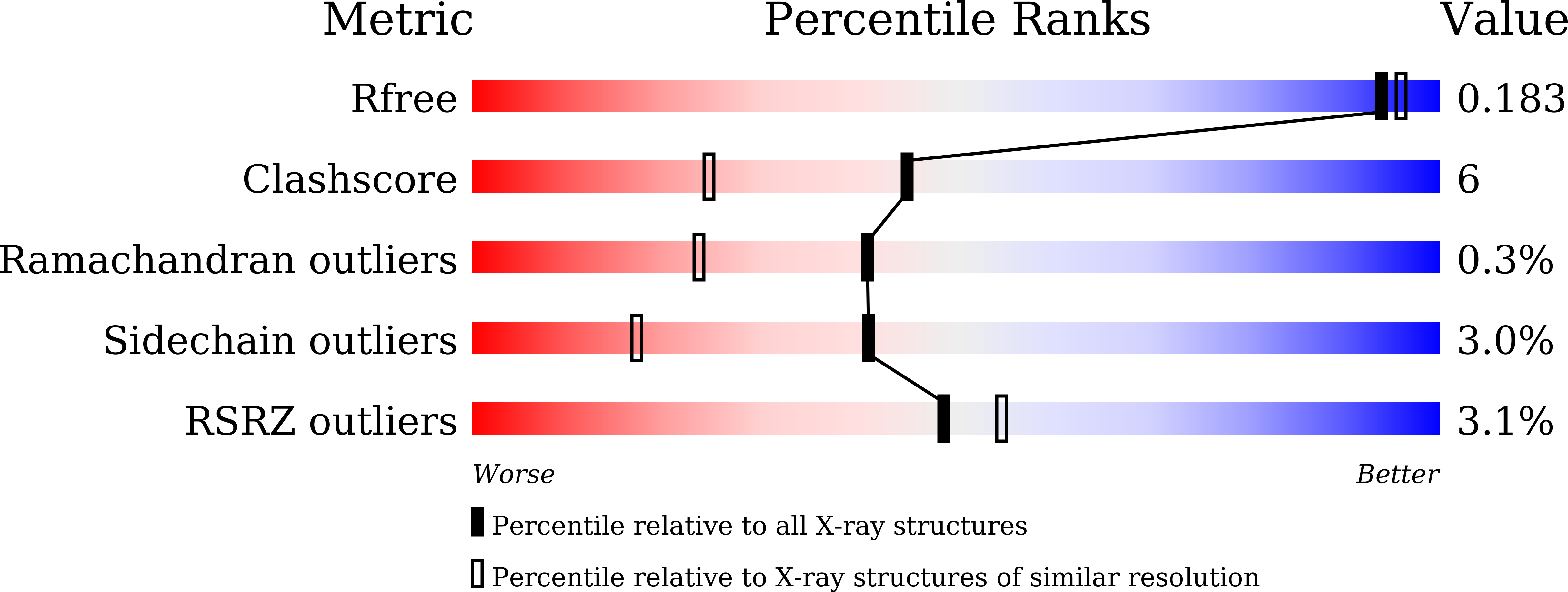
7D5X image
Entry Detail
PDB ID:
7D5X
Keywords:
Title:
Bovine heart cytochrome c oxidase in a catalytic intermediate, IO10, at 1.74 angstrom resolution
Biological Source:
Source Organism:
PDB Version:
Deposition Date:
2020-09-28
Release Date:
2021-07-21
Method Details:
Experimental Method:
Resolution:
1.74 Å
R-Value Free:
0.18
R-Value Work:
0.15
R-Value Observed:
0.15
Space Group:
P 21 21 21
Macromolecular Entities
Polymer Type:polypeptide(L)
Description:Cytochrome c oxidase subunit 1
Chain IDs:A, N
Chain Length:514
Number of Molecules:2
Biological Source:Bos taurus
Polymer Type:polypeptide(L)
Description:Cytochrome c oxidase subunit 2
Chain IDs:B, O
Chain Length:227
Number of Molecules:2
Biological Source:Bos taurus
Polymer Type:polypeptide(L)
Description:Cytochrome c oxidase subunit 3
Chain IDs:C, P
Chain Length:259
Number of Molecules:2
Biological Source:Bos taurus
Polymer Type:polypeptide(L)
Description:Cytochrome c oxidase subunit 4 isoform 1, mitochondrial
Chain IDs:D, Q
Chain Length:144
Number of Molecules:2
Biological Source:Bos taurus
Polymer Type:polypeptide(L)
Description:Cytochrome c oxidase subunit 5A, mitochondrial
Chain IDs:E, R
Chain Length:105
Number of Molecules:2
Biological Source:Bos taurus
Polymer Type:polypeptide(L)
Description:Cytochrome c oxidase subunit 5B, mitochondrial
Chain IDs:F, S
Chain Length:94
Number of Molecules:2
Biological Source:Bos taurus
Polymer Type:polypeptide(L)
Description:Cytochrome c oxidase subunit 6A2, mitochondrial
Chain IDs:G, T
Chain Length:84
Number of Molecules:2
Biological Source:Bos taurus
Polymer Type:polypeptide(L)
Description:Cytochrome c oxidase subunit 6B1
Chain IDs:H, U
Chain Length:79
Number of Molecules:2
Biological Source:Bos taurus
Polymer Type:polypeptide(L)
Description:Cytochrome c oxidase subunit 6C
Chain IDs:I, V
Chain Length:73
Number of Molecules:2
Biological Source:Bos taurus
Polymer Type:polypeptide(L)
Description:Cytochrome c oxidase subunit 7A1, mitochondrial
Chain IDs:J, W
Chain Length:58
Number of Molecules:2
Biological Source:Bos taurus
Polymer Type:polypeptide(L)
Description:Cytochrome c oxidase subunit 7B, mitochondrial
Chain IDs:K, X
Chain Length:49
Number of Molecules:2
Biological Source:Bos taurus
Polymer Type:polypeptide(L)
Description:Cytochrome c oxidase subunit 7C, mitochondrial
Chain IDs:L, Y
Chain Length:46
Number of Molecules:2
Biological Source:Bos taurus
Polymer Type:polypeptide(L)
Description:Cytochrome c oxidase subunit 8B, mitochondrial
Chain IDs:M, Z
Chain Length:43
Number of Molecules:2
Biological Source:Bos taurus
Modified Residue
Compound ID Chain ID Parent Comp ID Details 2D Image
FME A MET modified residue
SAC I SER modified residue
Primary Citation
Critical roles of the Cu B site in efficient proton pumping as revealed by crystal structures of mammalian cytochrome c oxidase catalytic intermediates.
J.Biol.Chem. 297 100967 100967 (2021)
PMID: 34274318 DOI: 10.1016/j.jbc.2021.100967

Abstact

Mammalian cytochrome c oxidase (CcO) reduces O2 to water in a bimetallic site including Fea3 and CuB giving intermediate molecules, termed A-, P-, F-, O-, E-, and R-forms. From the P-form on, each reaction step is driven by single-electron donations from cytochrome c coupled with the pumping of a single proton through the H-pathway, a proton-conducting pathway composed of a hydrogen-bond network and a water channel. The proton-gradient formed is utilized for ATP production by F-ATPase. For elucidation of the proton pumping mechanism, crystal structural determination of these intermediate forms is necessary. Here we report X-ray crystallographic analysis at ∼1.8 Å resolution of fully reduced CcO crystals treated with O2 for three different time periods. Our disentanglement of intermediate forms from crystals that were composed of multiple forms determined that these three crystallographic data sets contained ∼45% of the O-form structure, ∼45% of the E-form structure, and ∼20% of an oxymyoglobin-type structure consistent with the A-form, respectively. The O- and E-forms exhibit an unusually long CuB2+-OH- distance and CuB1+-H2O structure keeping Fea33+-OH- state, respectively, suggesting that the O- and E-forms have high electron affinities that cause the O→E and E→R transitions to be essentially irreversible and thus enable tightly coupled proton pumping. The water channel of the H-pathway is closed in the O- and E-forms and partially open in the R-form. These structures, together with those of the recently reported P- and F-forms, indicate that closure of the H-pathway water channel avoids back-leaking of protons for facilitating the effective proton pumping.

Legend

Protein

Chemical

Disease

Primary Citation of related structures