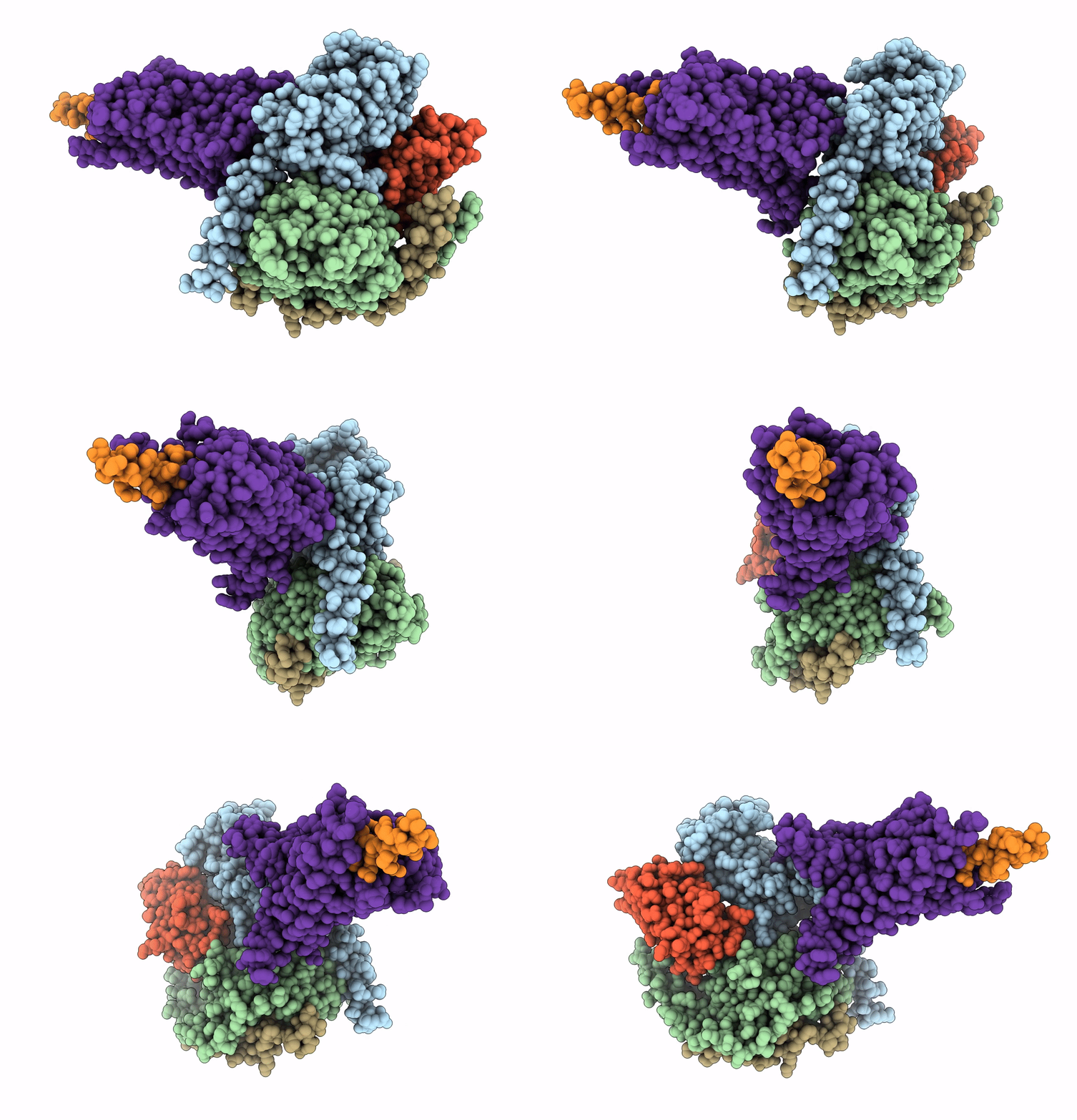
7D3S image
Deposition Date 2020-09-20
Release Date 2020-11-04
Last Version Date 2024-11-13
Entry Detail
PDB ID:
7D3S
Title:
Human SECR in complex with an engineered Gs heterotrimer
Biological Source:
Source Organism:
Homo sapiens (Taxon ID: 9606)
Rattus norvegicus (Taxon ID: 10116)
Bos taurus (Taxon ID: 9913)
unidentified (Taxon ID: 32644)
Host Organism:
Method Details:
Experimental Method:
Resolution:
2.90 Å
Aggregation State:
PARTICLE
Reconstruction Method:
SINGLE PARTICLE
Macromolecular Entities
Polymer Type:polypeptide(L)
Molecule:Guanine nucleotide-binding protein G(s) subunit alpha isoforms short
Gene (Uniprot):GNAS
Mutations:deletion 254-263, G49D,E50N,L63Y,A249D,S252D,I375A,V375I
Chain IDs:C (auth: A)
Chain Length:380
Number of Molecules:1
Biological Source:Homo sapiens
Polymer Type:polypeptide(L)
Molecule:Guanine nucleotide-binding protein G(I)/G(S)/G(T) subunit beta-1
Gene (Uniprot):Gnb1
Chain IDs:D (auth: B)
Chain Length:351
Number of Molecules:1
Biological Source:Rattus norvegicus
Polymer Type:polypeptide(L)
Molecule:Guanine nucleotide-binding protein G(I)/G(S)/G(O) subunit gamma-2
Gene (Uniprot):GNG2
Chain IDs:E (auth: G)
Chain Length:68
Number of Molecules:1
Biological Source:Bos taurus
Polymer Type:polypeptide(L)
Molecule:nanobody Nb35
Chain IDs:F (auth: N)
Chain Length:137
Number of Molecules:1
Biological Source:unidentified
Polymer Type:polypeptide(L)
Molecule:Secretin
Gene (Uniprot):SCT
Chain IDs:A (auth: P)
Chain Length:27
Number of Molecules:1
Biological Source:Homo sapiens
Polymer Type:polypeptide(L)
Molecule:Secretin receptor
Gene (Uniprot):SCTR
Chain IDs:B (auth: R)
Chain Length:434
Number of Molecules:1
Biological Source:Homo sapiens
Ligand Molecules
Primary Citation
Structure of the human secretin receptor coupled to an engineered heterotrimeric G protein.
Biochem.Biophys.Res.Commun. 533 861 866 (2020)
PMID: 33008599 DOI: 10.1016/j.bbrc.2020.08.042

Abstact

Secretin is a gastrointestinal hormone that exerts multiple physiological functions via activation of the secretin receptor (SECR). SECR belongs to the class B G-protein-coupled receptors and is involved in various processes, such as regulation of the pH of the duodenal content, food intake, and water homeostasis. Here, we report a cryo-electron microscopy structure of human SECR bound to secretin and an engineered Gs heterotrimer. The structure revealed the basic architecture of SECR and the secretin binding mode. A structural comparison of the SECR and PAC1R transmembrane domains revealed that transmembrane helices 1 and 2 play a prominent role in secretin recognition. Moreover, the extracellular domain of SECR is perpendicular to the TMD, unlike that of PAC1R. This comparison revealed the diverged peptide recognition mechanisms of these receptors, which belong to the same subgroup. Our structural information will facilitate drug discovery research for clinical applications.

Legend

Protein

Chemical

Disease

Primary Citation of related structures