7CXU image
Deposition Date 2020-09-02
Release Date 2021-07-14
Last Version Date 2023-11-29
Entry Detail
PDB ID:
7CXU
Title:
Crystal structure of CmnK in complex with NAD+
Biological Source:
Source Organism:
Method Details:
Experimental Method:
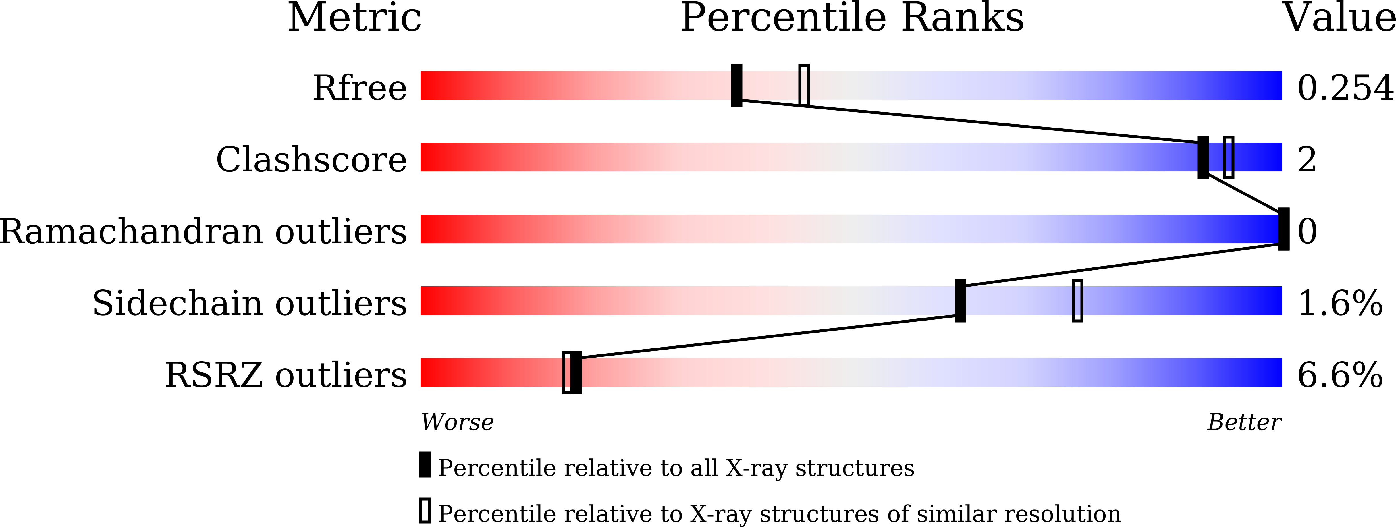
Resolution:
2.19 Å
R-Value Free:
0.24
R-Value Work:
0.20
R-Value Observed:
0.20
Space Group:
P 2 21 21
Macromolecular Entities
Structures with similar UniProt ID
Protein Blast
Polymer Type:polypeptide(L)
Molecule:CmnK
Gene (Uniprot):cmnK
Chain IDs:A, B
Chain Length:352
Number of Molecules:2
Biological Source:Saccharothrix mutabilis subsp. capreolus
Ligand Molecules
Primary Citation
Characterization of Enzymes Catalyzing the Formation of the Nonproteinogenic Amino Acid l-Dap in Capreomycin Biosynthesis.
Biochemistry 60 77 84 (2021)
PMID: 33356147 DOI: 10.1021/acs.biochem.0c00808

Abstact

Capreomycin (CMN) and viomycin (VIO) are nonribosomal peptide antituberculosis antibiotics, the structures of which contain four nonproteinogenic amino acids, including l-2,3-diaminopropionic acid (l-Dap), β-ureidodehydroalanine, l-capreomycidine, and β-lysine. Previous bioinformatics analysis suggested that CmnB/VioB and CmnK/VioK participate in the formation of l-Dap; however, the real substrates of these enzymes are yet to be confirmed. We herein show that starting from O-phospho-l-Ser (OPS) and l-Glu precursors, CmnB catalyzes the condensation reaction to generate a metabolite intermediate N-(1-amino-1-carboxyl-2-ethyl)glutamic acid (ACEGA), which undergoes NAD+-dependent oxidative hydrolysis by CmnK to generate l-Dap. Furthermore, the binding site of ACEGA and the catalytic mechanism of CmnK were elucidated with the assistance of three crystal structures, including those of apo-CmnK, the NAD+-CmnK complex, and CmnK in an alternative conformation. The CmnK-ACEGA docking model revealed that the glutamate α-hydrogen points toward the nicotinamide moiety. It provides evidence that the reaction is dependent on hydride transfer to form an imine intermediate, which is subsequently hydrolyzed by a water molecule to produce l-Dap. These findings modify the original proposed pathway and provide insights into l-Dap formation in the biosynthesis of other related natural products.

Legend

Protein

Chemical

Disease

Primary Citation of related structures