7CV0 image
Entry Detail
PDB ID:
7CV0
Title:
Crystal structure of B. halodurans NiaR in apo form
Biological Source:
PDB Version:
Deposition Date:
2020-08-25
Release Date:
2020-12-16
Method Details:
Experimental Method:
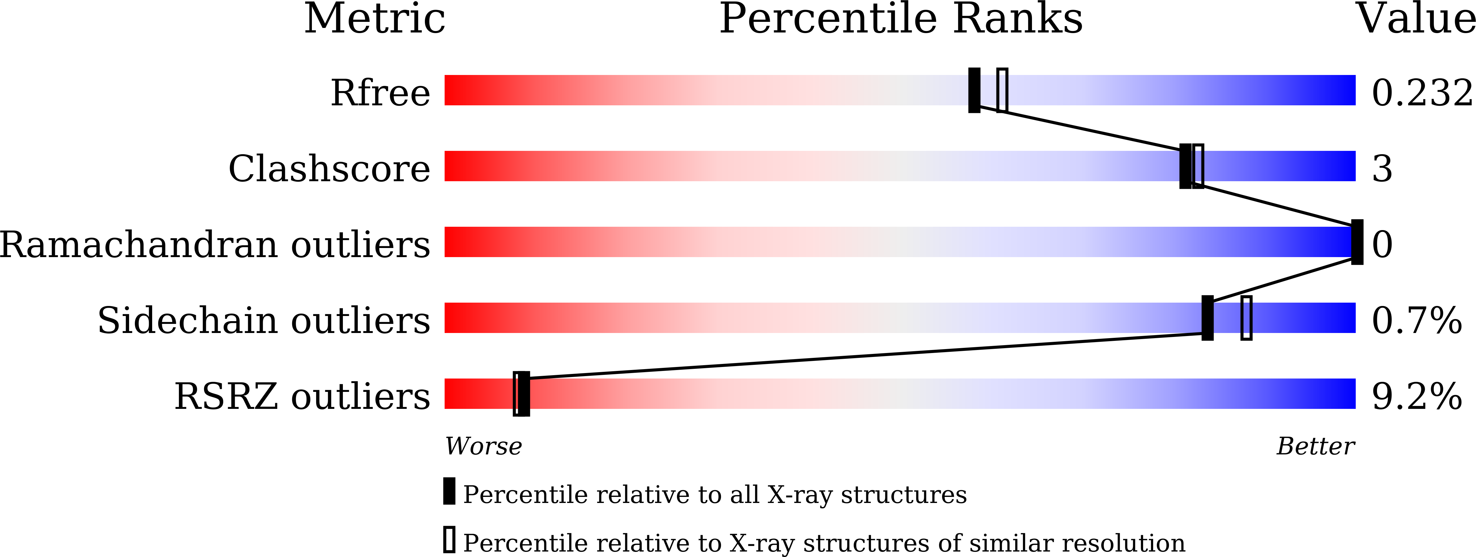
Resolution:
2.00 Å
R-Value Free:
0.22
R-Value Work:
0.20
R-Value Observed:
0.20
Space Group:
P 43 21 2
Macromolecular Entities
Polymer Type:polypeptide(L)
Description:Transcriptional regulator NiaR
Chain IDs:A
Chain Length:179
Number of Molecules:1
Biological Source:Bacillus halodurans (strain ATCC BAA-125 / DSM 18197 / FERM 7344 / JCM 9153 / C-125)
Ligand Molecules
Primary Citation
Structural analysis and insight into effector binding of the niacin-responsive repressor NiaR from Bacillus halodurans.
Sci Rep 10 21039 21039 (2020)
PMID: 33273654 DOI: 10.1038/s41598-020-78148-x

Abstact

The niacin-responsive repressor, NiaR, is transcriptional repressor of certain nicotinamide adenine dinucleotide (NAD) biosynthetic genes in response to an increase in niacin levels. NAD is a vital molecule involved in various cellular redox reactions as an electron donor or electron acceptor. The NiaR family is conserved broadly in the Bacillus/Clostridium group, as well as in the Fusobacteria and Thermotogales lineages. The NiaR structure consists of two domains: an N-terminal DNA-binding domain, and a C-terminal regulation domain containing a metal-binding site. In this paper, we report the crystal structures of apo and niacin-bound forms of NiaR from Bacillus halodurans (BhNiaR). The analysis of metal-binding and niacin-binding sites through the apo and niacin-bound structures is described. Each N- and C-terminal domain structure of BhNiaR is almost identical with NiaR from Thermotoga maritima, but the overall domain arrangement is quite different. A zinc ion is fully occupied in each subunit with well-conserved residues in the C-terminal domain. Niacin is also located at a hydrophobic pocket near the zinc ion in the C-terminal domain.

Legend

Protein

Chemical

Disease

Primary Citation of related structures