Deposition Date
2020-08-22
Release Date
2021-08-25
Last Version Date
2024-05-29
Entry Detail
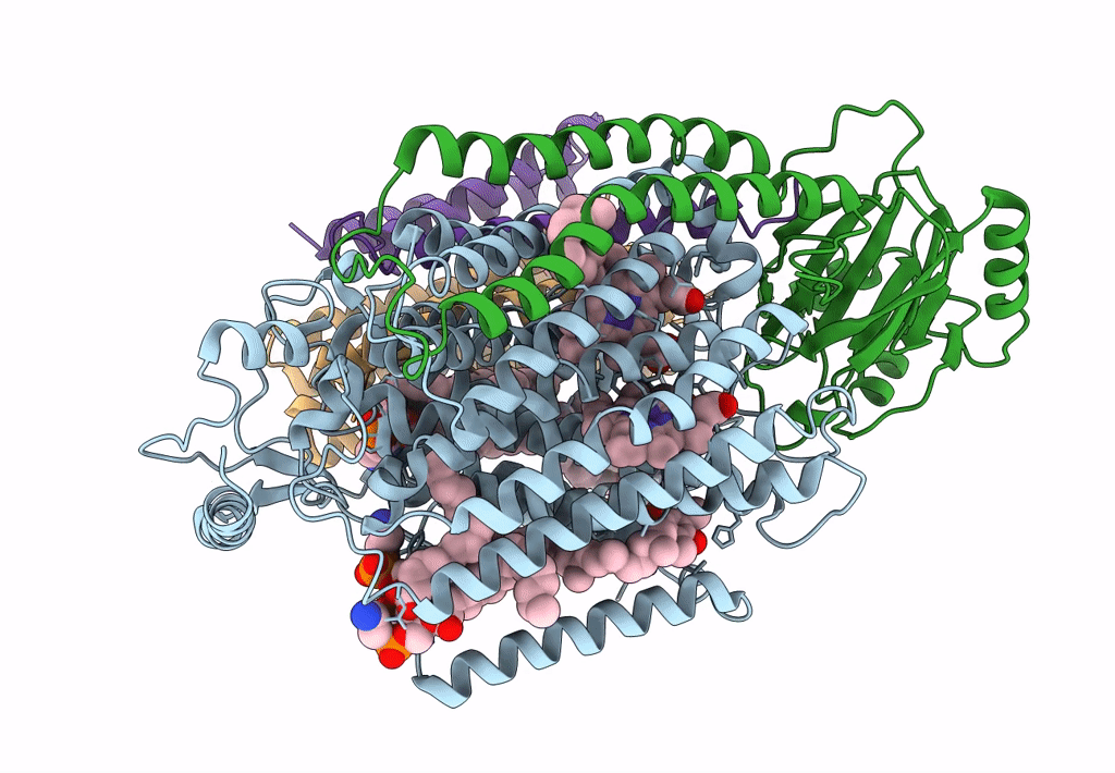
PDB ID:
7CUB
Keywords:
Title:
2.55-Angstrom Cryo-EM structure of Cytochrome bo3 from Escherichia coli in Native Membrane
Biological Source:
Source Organism(s):
Escherichia coli (Taxon ID: 562)
Method Details:
Experimental Method:
Resolution:
2.55 Å
Aggregation State:
PARTICLE
Reconstruction Method:
SINGLE PARTICLE