
Deposition Date
2020-08-18
Release Date
2021-03-03
Last Version Date
2023-11-29
Entry Detail
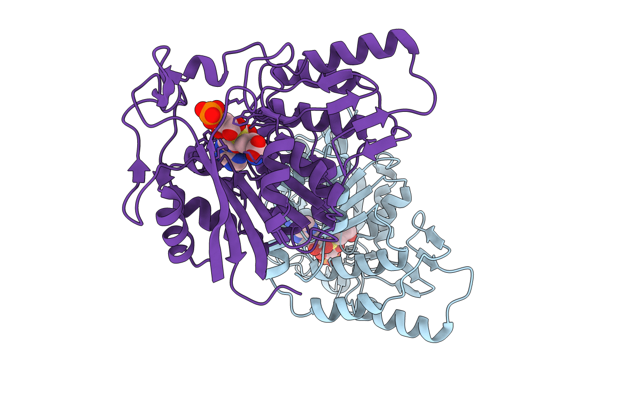
PDB ID:
7CT9
Keywords:
Title:
Crystal structure of SAH bound CmoB from Vibrio Vulnificus
Biological Source:
Source Organism(s):
Vibrio vulnificus MO6-24/O (Taxon ID: 914127)
Expression System(s):
Method Details:
Experimental Method:
Resolution:
2.30 Å
R-Value Free:
0.22
R-Value Work:
0.17
R-Value Observed:
0.17
Space Group:
C 1 2 1
