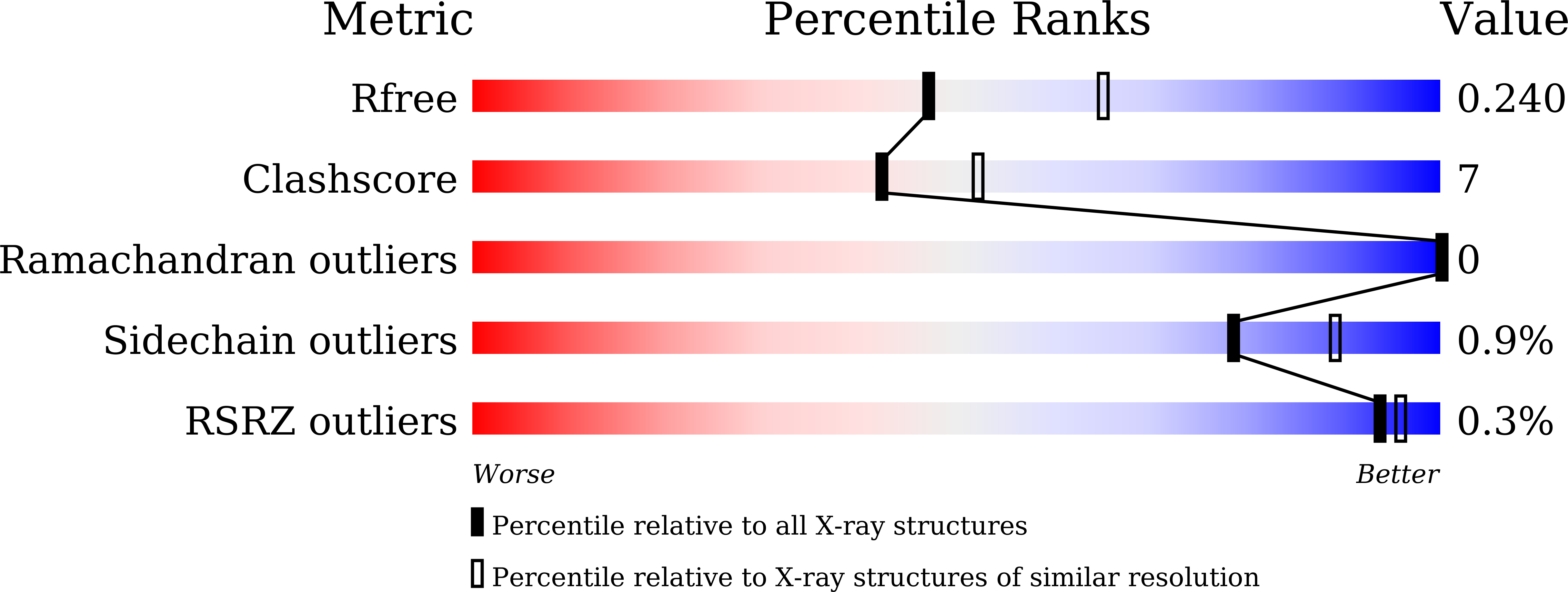
7CPH image
Deposition Date 2020-08-07
Release Date 2021-06-02
Last Version Date 2023-11-29
Entry Detail
PDB ID:
7CPH
Keywords:
Title:
Crystal structure of tRNA adenosine deaminase from Bacillus subtilis
Biological Source:
Method Details:
Experimental Method:
Resolution:
2.30 Å
R-Value Free:
0.23
R-Value Work:
0.17
R-Value Observed:
0.18
Space Group:
P 21 21 21
Macromolecular Entities
Structures with similar UniProt ID
Protein Blast
Polymer Type:polypeptide(L)
Molecule:tRNA-specific adenosine deaminase
Gene (Uniprot):tadA
Chain IDs:A, B
Chain Length:167
Number of Molecules:2
Biological Source:Bacillus subtilis subsp. subtilis str. 168
Primary Citation
Deciphering protein microenvironment by using a cysteine specific switch-ON fluorescent probe.
Org.Biomol.Chem. 19 5161 5168 (2021)
PMID: 34037063 DOI: 10.1039/d1ob00698c

Abstact

Fluorescent probes provide an unparalleled opportunity to visualize and quantify dynamic events. Here, we employ a medium-size, cysteine specific coumarin based switch-ON fluorescent probe 'L' to track protein unfolding profiles and accessibility of cysteine residues in proteins. It was established that 'L' is highly selective and exhibits no artifact due to interaction with other bystander species. 'L' is able to gauge subtle changes in protein microenvironment and proved to be effective in delineating early unfolding events that are difficult to otherwise discern by classic techniques such as circular dichroism. By solving the X-ray structure of TadA and probing the temperature dependent fluorescence-ON response with native TadA and its cysteine mutants, it was revealed that unfolding occurs in a stage-wise manner and the regions that are functionally important form compact sub-domains and unfold at later stages. Our results assert that probe 'L' serves as an efficient tool to monitor subtle changes in protein structure and can be employed as a generic dye to study processes such as protein unfolding.

Legend

Protein

Chemical

Disease

Primary Citation of related structures