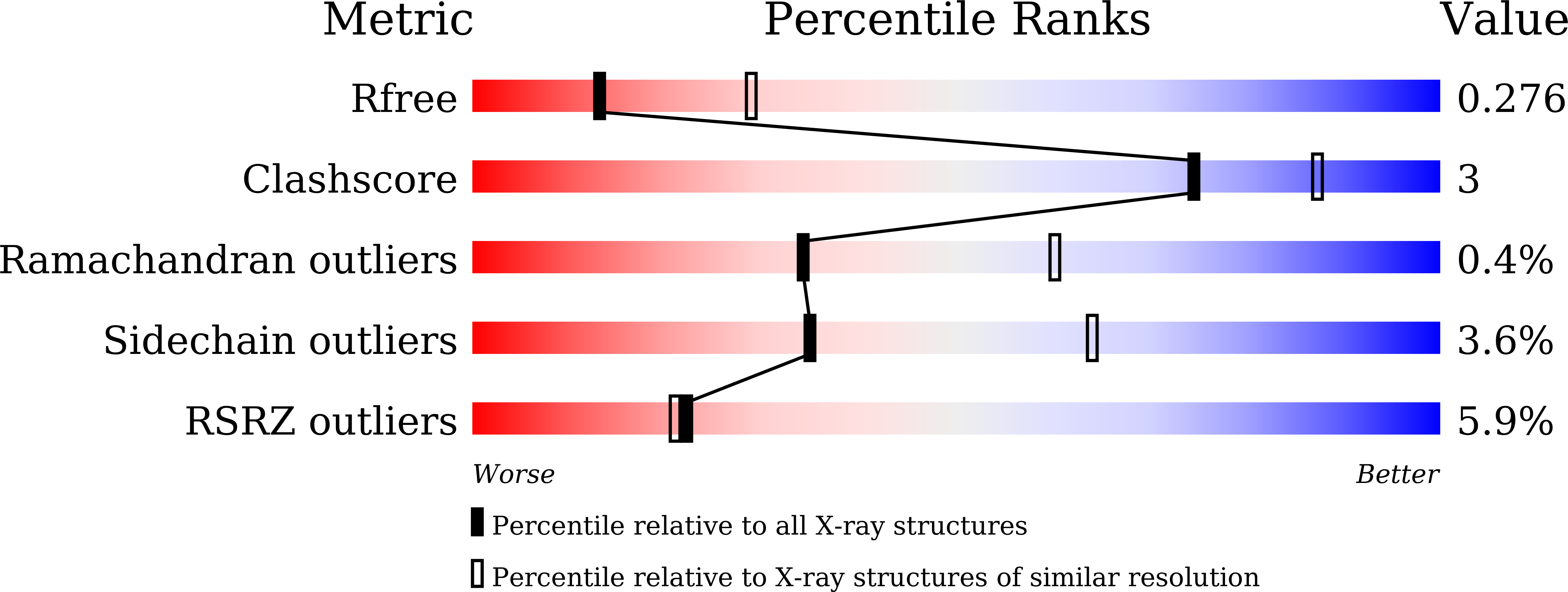
7CNZ image
Entry Detail
PDB ID:
7CNZ
Keywords:
Title:
Crystal structure of 10PE bound PSD from E. coli (2.70 A)
Biological Source:
Source Organism:
PDB Version:
Deposition Date:
2020-08-03
Release Date:
2021-03-24
Method Details:
Experimental Method:
Resolution:
2.70 Å
R-Value Free:
0.27
R-Value Work:
0.22
R-Value Observed:
0.22
Space Group:
P 21 21 21
Macromolecular Entities
Polymer Type:polypeptide(L)
Description:Phosphatidylserine decarboxylase beta chain
Chain IDs:A, C, E, G
Chain Length:253
Number of Molecules:4
Biological Source:Escherichia coli K-12
Polymer Type:polypeptide(L)
Description:Phosphatidylserine decarboxylase alpha chain
Chain IDs:B, D, F, H
Chain Length:42
Number of Molecules:4
Biological Source:Escherichia coli K-12
Primary Citation
Structural insights into phosphatidylethanolamine formation in bacterial membrane biogenesis.
Sci Rep 11 5785 5785 (2021)
PMID: 33707636 DOI: 10.1038/s41598-021-85195-5

Abstact

Phosphatidylethanolamine (PE), a major component of the cellular membrane across all domains of life, is synthesized exclusively by membrane-anchored phosphatidylserine decarboxylase (PSD) in most bacteria. The enzyme undergoes auto-cleavage for activation and utilizes the pyruvoyl moiety to form a Schiff base intermediate with PS to facilitate decarboxylation. However, the structural basis for self-maturation, PS binding, and decarboxylation processes directed by PSD remain unclear. Here, we present X-ray crystal structures of PSD from Escherichia coli, representing an apo form and a PE-bound complex, in which the phospholipid is chemically conjugated to the essential pyruvoyl residue, mimicking the Schiff base intermediate. The high-resolution structures of PE-complexed PSD clearly illustrate extensive hydrophobic interactions with the fatty acyl chains of the phospholipid, providing insights into the broad specificity of the enzyme over a wide range of cellular PS. Furthermore, these structures strongly advocate the unique topology of the enzyme in a lipid bilayer environment, where the enzyme associates with cell membranes in a monotopic fashion via the N-terminal domain composed of three amphipathic helices. Lastly, mutagenesis analyses reveal that E. coli PSD primarily employs D90/D142-H144-S254 to achieve auto-cleavage for the proenzyme maturation, where D90 and D142 act in complementary to each other.

Legend

Protein

Chemical

Disease

Primary Citation of related structures