Deposition Date
2020-07-20
Release Date
2020-10-28
Last Version Date
2023-11-29
Entry Detail
PDB ID:
7CLA
Keywords:
Title:
Crystal structure of HTH-type transcriptional regulator SkgA from Caulobacter crescentus
Biological Source:
Source Organism(s):
Expression System(s):
Method Details:
Experimental Method:
Resolution:
2.50 Å
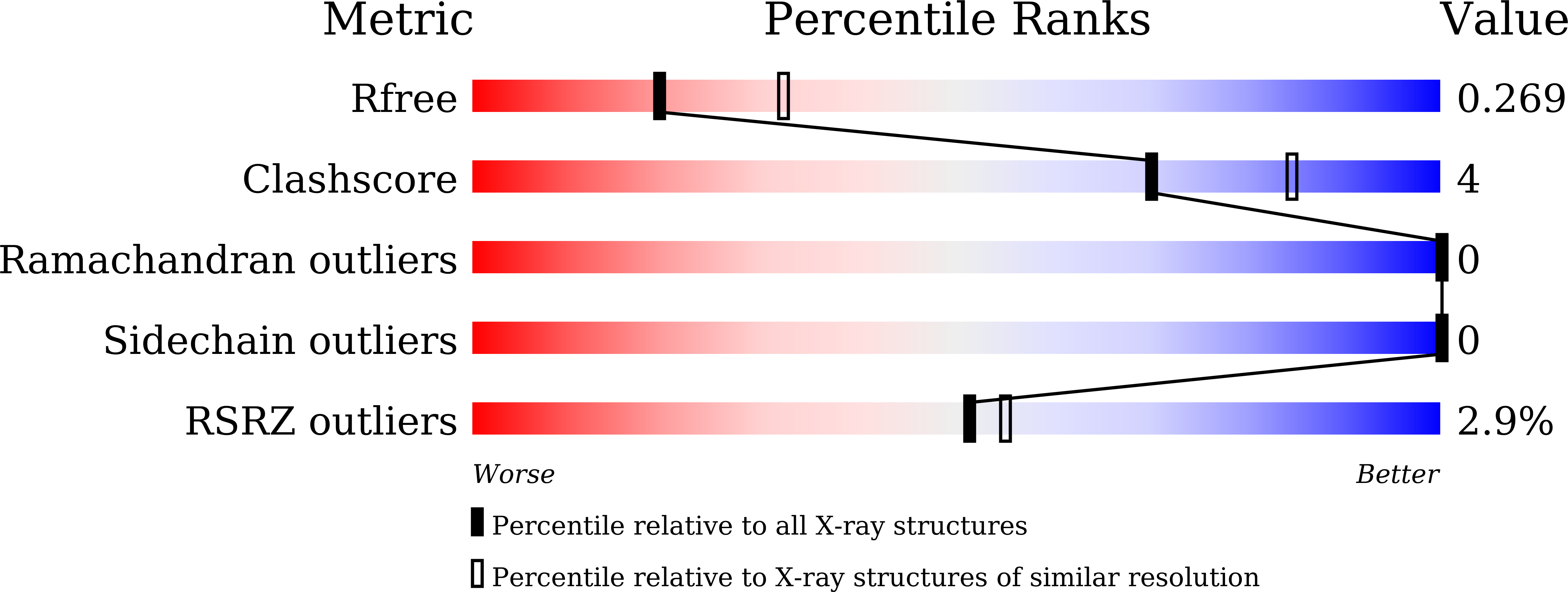
R-Value Free:
0.26
R-Value Work:
0.20
R-Value Observed:
0.21
Space Group:
P 65 2 2