7CK5 image
Deposition Date 2020-07-15
Release Date 2021-07-21
Last Version Date 2024-05-15
Entry Detail
PDB ID:
7CK5
Keywords:
Title:
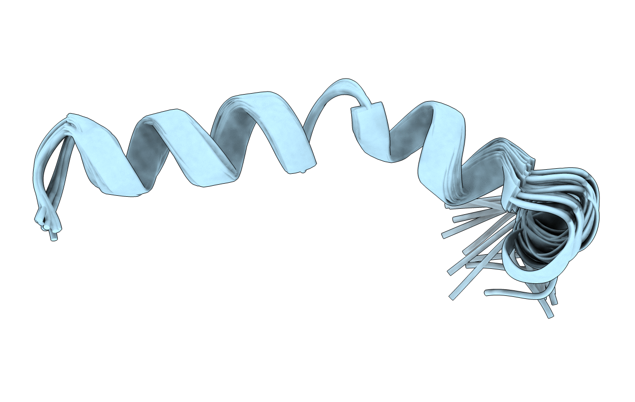
Solution structure of 28 amino acid polypeptide (354-381) in Plantago asiatica mosaic virus replicase bound to SDS micelle
Biological Source:
Source Organism:
Host Organism:
Method Details:
Experimental Method:
Conformers Calculated:
100
Conformers Submitted:
20
Selection Criteria:
structures with the lowest energy
Macromolecular Entities
Structures with similar UniProt ID
Protein Blast
Polymer Type:polypeptide(L)
Molecule:PlAMV replicase peptide from RNA-dependent RNA polymerase
Gene (Uniprot):RdRp
Chain IDs:A
Chain Length:28
Number of Molecules:1
Biological Source:Plantago asiatica mosaic potexvirus
Ligand Molecules
Primary Citation
Identification of a Proline-Kinked Amphipathic alpha-Helix Downstream from the Methyltransferase Domain of a Potexvirus Replicase and Its Role in Virus Replication and Perinuclear Complex Formation.
J.Virol. 95 e0190620 e0190620 (2021)
PMID: 34346768 DOI: 10.1128/JVI.01906-20

Abstact

Characterized positive-strand RNA viruses replicate in association with intracellular membranes. Regarding viruses in the genus Potexvirus, the mechanism by which their RNA-dependent RNA polymerase (replicase) associates with membranes is understudied. Here, by membrane flotation analyses of the replicase of Plantago asiatica mosaic potexvirus (PlAMV), we identified a region in the methyltransferase (MET) domain as a membrane association determinant. An amphipathic α-helix was predicted downstream from the core region of the MET domain, and hydrophobic amino acid residues were conserved in the helical sequences in replicases of other potexviruses. Nuclear magnetic resonance (NMR) analysis confirmed the amphipathic α-helical configuration and unveiled a kink caused by a highly conserved proline residue in the α-helix. Substitution of this proline residue and other hydrophobic and charged residues in the amphipathic α-helix abolished PlAMV replication. Ectopic expression of a green fluorescent protein (GFP) fusion with the entire MET domain resulted in the formation of a large perinuclear complex, where virus replicase and RNA colocated during virus infection. Except for the proline substitution, the amino acid substitutions in the α-helix that abolished virus replication also prevented the formation of the large perinuclear complex by the respective GFP-MET fusion. Small intracellular punctate structures were observed for all GFP-MET fusions, and in vitro high-molecular-weight complexes were formed by both replication-competent and -incompetent viral replicons and thus were not sufficient for replication competence. We discuss the roles of the potexvirus-specific, proline-kinked amphipathic helical structure in virus replication and intracellular large complex and punctate structure formation. IMPORTANCE RNA viruses characteristically associate with intracellular membranes during replication. Although virus replicases are assumed to possess membrane-targeting properties, their membrane association domains generally remain unidentified or poorly characterized. Here, we identified a proline-kinked amphipathic α-helix structure downstream from the methyltransferase core domain of PlAMV replicase as a membrane association determinant. This helical sequence, which includes the proline residue, was conserved among potexviruses and related viruses in the order Tymovirales. Substitution of the proline residue, but not the other residues necessary for replication, allowed formation of a large perinuclear complex within cells resembling those formed by PlAMV replicase and RNA during virus replication. Our results demonstrate the role of the amphipathic α-helix in PlAMV replicase in a perinuclear complex formation and virus replication and that perinuclear complex formation by the replicase alone will not necessarily indicate successful virus replication.

Legend

Protein

Chemical

Disease

Primary Citation of related structures