Deposition Date
2020-07-08
Release Date
2020-12-09
Last Version Date
2024-10-30
Entry Detail
PDB ID:
7CIO
Keywords:
Title:
Molecular interactions of cytoplasmic region of CTLA-4 with SH2 domains of PI3-kinase
Biological Source:
Source Organism(s):
Homo sapiens (Taxon ID: 9606)
Expression System(s):
Method Details:
Experimental Method:
Resolution:
1.10 Å
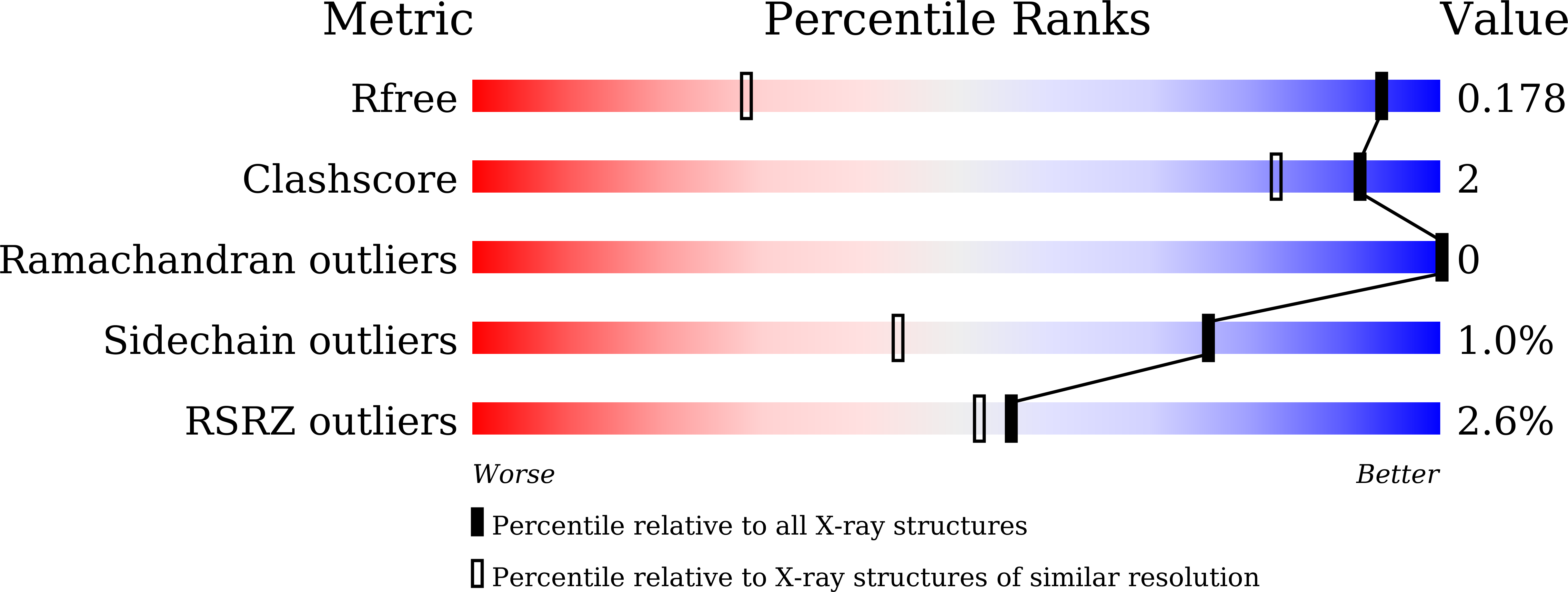
R-Value Free:
0.17
R-Value Work:
0.15
R-Value Observed:
0.15
Space Group:
P 21 21 21